
Abstract
Objective
Osteosarcoma is one of the most common malignancies in children and adolescents. Studies have shown that miR-34c-5p is involved in the progression of various cancers. To explore the effects of miR-34c-5p on the proliferation, migration and invasion of osteosarcoma cells and its potential mechanism.
Methods
qRT-PCR was used to detect the expression levels of miR-34c-5p and FLOT2 mRNA in osteosarcoma tissues and cells. Western Blot was used to detect protein expression. MTT assay used to detect cell viability. Transwell was used to detect cell migration and invasion in each group. Dual luciferase reporter gene assay was used to detect luciferase activity.
Results
The expression of miR-34c-5pwas significantly decreased in osteosarcoma tissues and cells and the expression level of FLOT2 mRNA was significantly increased. Overexpression of miR-34c-5p and inhibition of FLOT2 inhibited the proliferation, migration and invasion of osteosarcoma cells and inhibited the expression of Cyclin D1, MMP-2 and MMP-9 proteins and promoted the expression of p21 protein. miR-34c-5p targeted to regulate the expression of FLOT2. Overexpression of FLOT2 reversed the inhibitory effect of miR-34c-5p overexpression on proliferation, migration and invasion of osteosarcoma cell lines.
Conclusion
miR-34c-5p can inhibit the proliferation, migration and invasion of osteosarcoma cells. The mechanism may be related to targeting FLOT2, which will provide a new target for the prevention and treatment of osteosarcoma.
Introduction
Osteosarcoma (OS) is a malignant bone tumor commonly seen in adolescents and children. At present, surgery combined with systemic chemotherapy is still the main treatment. Although the survival rate is not very low, clinical osteosarcoma is prone to recurrence, metastasis and drug resistance, high disability and mortality, poor prognosis, and new treatment methods and treatment targets are urgently needed [Citation1]. It has been found that the abnormal expression of many miRNAs in osteosarcoma is closely related to its occurrence, metastasis and drug resistance. It can be used as a biomarker for the diagnosis and treatment of osteosarcoma [Citation2]. miR-34c-5p is involved in the progression of various tumors, such as miR-34c-5p enhances the sensitivity of cervical cancer cells to pirarubicin [Citation3]; miR-34c-5p is underexpressed in gastric cancer and overexpression significantly increases the chemosensitivity of paclitaxel-resistant gastric cancer cells [Citation4]. miR-34c-5p methylation inhibits proliferation and metastasis of colorectal cancer and attenuates epithelial-mesenchymal transition [Citation5]. miR-34c-5p is under-expressed in laryngeal squamous cell carcinoma and is associated with prognosis [Citation6]. miR-34c-5p promotes HIV-1 activation and replication [Citation7]. miR-34c-5p can inhibit the proliferation, migration and invasion of SiHa cells and promote apoptosis [Citation8]. miR-34c-5p is under-expressed in nasopharyngeal carcinoma and its expression is up-regulated to inhibit the proliferation and metastasis of nasopharyngeal carcinoma cells [Citation9]. However, the potential molecular mechanism of miR-34c-5p in osteosarcoma has not been studied.
Flotillin-2 (FLOT2) is an important protein of planar lipid valve on the cell membrane. It is abnormally expressed in nasopharyngeal carcinoma, breast cancer, gastric cancer, renal cancer, malignant melanoma and other tumors, which is closely related to the proliferation, invasion and metastasis of tumors [Citation10]. This study was to investigate the effect of miR-34c-5p on proliferation, migration and invasion of osteosarcoma cells and its relationship with FLOT2, which will provide a new target for targeted therapy of osteosarcoma.
Materials and methods
Materials
Human osteosarcoma cell lines MG63, U-2OS, SAOS-2 and human normal osteoblast cell line hFOB1.19 were purchased from ATCC (American Type Culture Collection, Manassas, VA, USA). Fetal bovine serum, RPMI-1640 medium, trypsin was purchased from Gibico (Grand Island, NY, USA). LipofectamineTM 2000 transfection reagent was purchased from Invitrogen, USA. MTT kit and dual luciferase reporter gene detection kit were purchased from Beijing Solarbio (Beijing, China). Trizol reagent, reverse transcription kit, and fluorescence quantitative kit were purchased from TaKaRa (Tokyo, Japan). Transwell chamber and Matrigel gel was purchased from BD Biosciences (Franklin Lakes, New Jersey, USA). Dimethyl sulfoxide (DMSO), BCA kit, and PBS buffer were purchased from Sigma-Aldrich (St. Louis, MO,USA). RIPA protein lysate and SDS-PAGE kit was purchased from Shanghai Biyuntian Biotechnology Co., Ltd. (Shanghai, China).
Methods
Tissue samples
The 20 pairs of osteosarcoma tissues and adjacent normal tissues were collected from Armed Police Medical Center Hospital. All tissues samples were stored at −80 °C after quick freezing in liquid nitrogen. Written informed consent was obtained from all participants. The research method was approved by the Ethics Committee of Armed Police Medical Center hospital.
Cell culture
Human osteosarcoma cells MG63, U-2OS, SAOS-2 and human normal osteoblasts hFOB1.19 were cultured in RPMI-1640 medium containing 10% fetal bovine serum at 37 °C, 5% CO2 saturated humidity, and changed once a day. When the cells are fused to about 70%, trypsin was added for digestion and passage and cells in the logarithmic growth phase were selected for experiments.
Cell transfection and grouping
Conventional cultured human osteosarcoma cells MG63 and SAOS-2 were digested with 0.25% trypsin and seeded in 96-well plates. After the cells were grown to 80% confluence, they were replaced with serum-free medium for 12 h. Then, miR-NC, miR-34c-5p, si-NC, si-FLOT2, anti-miR-NC or anti-miR-34c- 5p was transfected into osteosarcoma cell lines MG63 and SAOS-2, which was recorded as miR-NC group, miR-34c-5p group, si-NC group, si-FLOT2 group, anti-miR-NC group or anti-miR-34c-5p group. The miR-34c-5p and pcDNA or pcDNA-FLOT2 plasmids were cotransfected into osteosarcoma cell lines MG63 and SAOS-2 and recorded as miR-34c-5p + pcDNA group or miR-34c-5p + pcDNA-FLOT2 group. Transfections were performed according to the LipofectamineTM 2000 kit (Invitrogen, Carlsbad, CA, USA).
MTT detection of cell proliferation
In the miR-NC group, miR-34c-5p group, si-NC group, si-FLOT2 group, miR-34c-5p + pcDNA group and miR-34c-5p + pcDNA-FLOT2 group, the cells were cultured for 24 h, 48 h and 72 h. Added 20 μl (5 g/L) MTT solution and continue to incubate for 4 h. The excess medium was discarded and reacted with 150 μl DMSO for 10 min. The absorbance (A) at 490 nm was measured by a microplate reader. Cell proliferation activity (%) = experimental group A value/blank control group A value × 100%.
Transwell detection of cell migration and invasion
miR-NC group, miR-34c-5p group, si-NC group, si-FLOT2 group, miR-34c-5p + pcDNA group and miR-34c-5p + pcDNA-FLOT2 group cells were re-suspended in the serum-free medium after trypsinase digestion and the concentration was adjusted to 2 × 10⁴ cells/ml.
Cell migration assay
200 μl cell suspension was inoculated into the upper chamber of Transwell chamber and placed in the lower chamber containing complete medium, and cultured at 37 °C, 5% CO2 for 24 h. After removing the culture medium, the upper cells were gently wiped with a cotton swab. The lower cells were fixed with 4% paraformaldehyde for 30 min and stained with 0.1% crystal violet for 10 min, and observed under a microscope and took photos randomly in five visual fields. The number of cells stained with crystal violet was the number of migrating cells.
Cell invasion experiment
After diluting Matrigel with RPMI-1640 medium at a ratio of 1:5, it was laid in the upper chamber of the Transwell chamber. After drying at room temperature, it was operated according to the steps of cell migration experiment. Finally, the number of cells stained with crystal violet was observed under the microscope.
qRT-PCR detection of miR-34c-5p and FLOT2 mRNA expression levels
Cells in each group were collected and ground sufficiently. Total RNA was extracted by Trizol reagent. RNA purity was detected by micronucleic acid analyzer. The RNA was reversely transcribed into cDNA using the TaKaRa reverse transcription kit. The reaction system was prepared according to the TaKaRa fluorescence quantification kit. PCR amplification was performed with β-actin as internal reference and each sample was repeated 3 times. The cycling conditions were 95 °C for 30 s, 60 °C for 30 s, 72 °C for 30 s for 40 cycles, and 60 °C for 5 min. The relative expression amount was calculated by the 2−ΔΔCt method.
Western Blot detection of protein expression
The cells of each group were collected and lysed by RIPA lysate and then were centrifuged at 12,000 g for 15 min at 4 °C. Protein supernatant was collected and protein concentration was determined by BCA Kit (Thermo Scientific, Grand Island, NY, USA). The protein samples were subjected to SDS-PAGE electrophoresis and transferred to a PVDF membrane. Then the 5% skim milk powder blocking solution was blocked at room temperature for 1 h. Added primary antibody (1:1000), and incubated at 4 °C overnight and washed the membrane with PBS. Added secondary antibody (1:2000) for 2 h at room temperature. The membrane was washed 3 times with TBST for 10 min each time, then exposed and developed in dark room, then immersed and fixed, and finally, the residue was washed and dried. The film was processed with Quantity One gel analysis software (Bio-Rad Laboratories, Hercules, CA, USA) and the ratio of the target band to the GAPDH band was used as the protein expression level. Each protein sample was repeated 3 times.
Luciferase reporter gene assay to detect the targeted regulation of FLOT2 by miR-34c-5p
The TargetScan database showed that the FLOT2 3'UTR region had a miR-34c-5p binding site. Osteosarcoma cells MG63 and SAOS-2 in logarithmic growth phase were seeded in 24-well plates (5 × 10⁴ cells/well). When cells were grown to 80% confluence, the pGL3 plasmid harboring the wild-type or mutant FLOT2 3′UTR was cotransfected with miR-NC or miR-34c-5p into osteosarcoma cells using LipofectamineTM 2000 (Invitrogen, Carlsbad, CA, USA). According to the requirements of the instructions, dual luciferase reporter gene detection was used to detect dual luciferase reporter gene. The results were analyzed by the ratio of luciferase activity to Renilla activity. The experiment was repeated three times.
Statistical analysis
Statistical analysis was performed using SPSS 20.0 (SPSS Inc., Chicago, IL, USA). The measurement data were expressed as mean ± standard deviation. The two groups were compared by t-test. The comparison among groups was analyzed by one-way ANOVA. The difference was statistically significant at p < .05.
Results
Expression of miR-34c-5p and FLOT2 in osteosarcoma tissues and cells
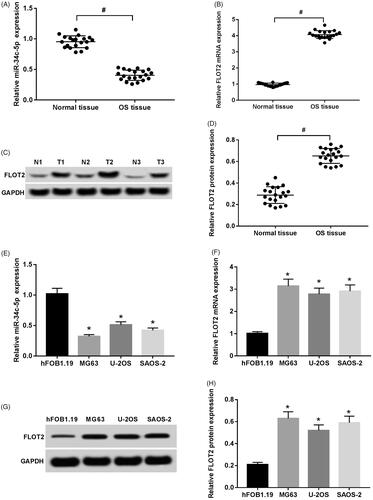
Firstly, qRT-PCR assay revealed that miR-34c-5p was significantly down-regulated in osteosarcoma tissues compared to adjacent normal tissues (), while FLOT2 was strikingly upregulated () (p < .05). In addition, Western Blot assay suggested that FLOT2 protein expression in osteosarcoma tissues was increased compared with adjacent normal tissues (). The results of qRT-PCR assay () showed that the expression level of miR-34c-5p in human osteosarcoma cells MG63, U-2OS and SAOS-2 were significantly lower than those of human normal osteoblasts hFOB1.19. The expression level of FLOT2 mRNA was significantly increased (p < .05). The results of Western blot assay () showed that the expression level of FLOT2 protein in human osteosarcoma cells MG63, U-2OS and SAOS-2 was significantly higher than that of human normal osteoblast hFOB1.19 (p < .05).
Figure 1. Expression of miR-34c-5p and FLOT2 in osteosarcoma tissues and cells. (A) The expression of miR-34c-5p in osteosarcoma tissues and adjacent normal tissues. (B) The expression of FLOT2 mRNA in osteosarcoma tissues and adjacent normal tissues. (C, D) The expression of FLOT2 protein in osteosarcoma tissues and adjacent normal tissues. (E) Expression of miR-34c-5p in osteosarcoma cells and normal osteoblasts. (F) Expression of FLOT2 mRNA in osteosarcoma cells and normal osteoblasts. (G, H) Expression of FLOT2 protein in osteosarcoma cells and normal osteoblasts. Note: Compared with hFOB1.19 group, #p < .01, *p < .05 (B–D).
Effect of miR-34c-5p overexpression on proliferation of osteosarcoma cells
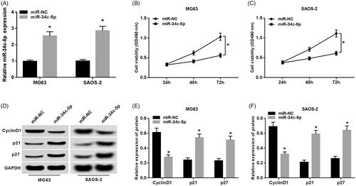
The results of qRT-PCR assay () showed that the expression level of miR-34c-5p was significantly increased in the miR-34c-5p group compared with the miR-NC group (p < .05). The MTT assay results () showed that cell viability was measured at 24 h, 48 h and 72 h after transfection of miR-NC or miR-34c-5p. Compared with the miR-NC group, the activity of osteosarcoma cells in the miR-34c-5p group was significantly decreased (p < .05). The results of Western Blot () showed that the expression level of CyclinD1 protein in MG63 and SAOS-2 cells of the miR-34c-5p group was significantly decreased compared with the miR-NC group, and the expression levels of p21 and p27 proteins were significantly increased (p < .05). It can be seen that miR-34c-5p overexpression inhibits the proliferation of osteosarcoma cells.
Figure 2. Effect of miR-34c-5p overexpression on proliferation of osteosarcoma cells. (A) Relative expression of miR-34c-5p. (B, C) Detection of osteosarcoma cell viability. (D–F) Expression of cell proliferation-related proteins. Note: Compared with miR-NC group, *p < .05.
Effect of miR-34c-5p overexpression on migration and invasion of osteosarcoma cells
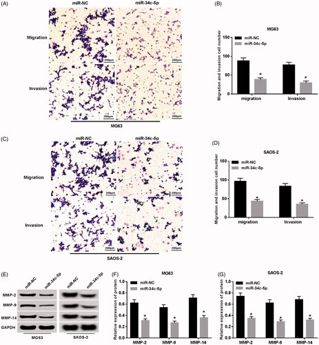
The results of the Transwell assay () showed that the number of migration and invasion of MG63 and SAOS-2 cells in the miR-34c-5p group was significantly decreased compared with the miR-NC group (p < .05). The results of Western Blot () showed that the expression levels of MMP-2, MMP-9 and MMP-14 in MG63 and SAOS-2 cells of the miR-34c-5p group were significantly lower than those in the miR-NC group (p < .05). It can be seen that miR-34c-5p overexpression inhibits migration and invasion of osteosarcoma cells.
Figure 3. Effect of miR-34c-5p overexpression on migration and invasion of osteosarcoma cells. (A–D) The migration and invasion of osteosarcoma cells. (E–G) Expression of cell migration and invasion related proteins. Note: Compared with miR-NC group, *p < .05.
Effects of inhibition of FLOT2 expression on proliferation, migration and invasion of osteosarcoma cells
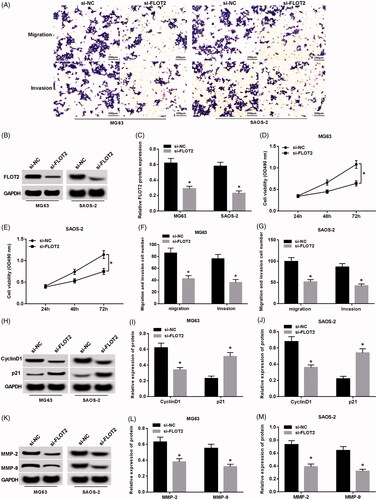
Western Blot results () showed that the expression level of FLOT2 protein was significantly lower in the si-FLOT2 group compared with the si-NC group. The MTT assay results () showed that cell viability was measured at 24 h, 48 h, and 72 h after transfection of si-NC or si-FLOT2. Compared with the si-NC group, the activity of MG63 and SAOS-2 cells in the si-FLOT2 group was significantly decreased (p < .05). The results of the Transwell assay () showed that the number of migration and invasion of MG63 and SAOS-2 cells in the si-FLOT2 group was significantly decreased compared with the si-NC group (p < .05). The proliferation-related protein CyclinD1 was remarkedly decreased in the si-FLOT2 group compared to the si-NC group, while the expression of p21 protein was significantly increased () (p < .05). Furthermore, the cell migration and invasion-related protein (MMP-2 and MMP-9) was markedly lower in the si-FLOT2 group compared with the si-NC group () (p < .05). It can be seen that inhibition of FLOT2 expression inhibits proliferation, migration and invasion of osteosarcoma cells.
Figure 4. Effect of inhibition of FLOT2 expression on proliferation, migration and invasion of osteosarcoma cells. (A) The migration and invasion of osteosarcoma cells. (B, C) Relative expression of FLOT2 protein. (D, E) Effect of inhibiting FLOT2 expression on proliferation of osteosarcoma cells. (F, G) Inhibition of FLOT2 expression on migration and invasion of osteosarcoma cells. (H–J) Effect of inhibition of FLOT2 expression on proliferation-related protein expression in osteosarcoma cells. (K–M) Effect of inhibition of FLOT2 expression on expression of migration-invasive-related proteins in osteosarcoma cells. Note: Compared with si-NC group, *p < .05
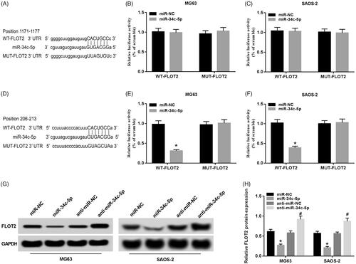
miR-34c-5p targets and regulates the expression of FLOT2
Binding sites for FLOT2 and miR-34c-5p was predicted by the TargetScan database (). The results of the luciferase reporter gene assay () showed that the luciferase activity of osteosarcoma cells transfected with wild-type FLOT2 gene expression vector WT-FLOT2 was significantly lower in miR-34c-5p group than that in miR-NC group (p < .05). After transfecting the mutant FLOT2 gene expression vector MUT-FLOT2, the luciferase activity of osteosarcoma cells in the miR-34c-5p group was not significantly different compared with the miR-NC group. However, luciferase reporter assay revealed that another binding site at 1171–1177 position had no effect on luciferase activity (). The results of Western Blot assay () showed that the expression level of FLOT2 protein in osteosarcoma cells of miR-34c-5p group was significantly lower than that of miR-NC group. Compared with the anti-NC group, the expression level of FLOT2 protein in osteosarcoma cells of anti-miR-34c-5p group was significantly increased (p < .05). It can be seen that miR-34c-5p can specifically regulate the expression of FLOT2.
Figure 5. miR-34c-5p targeted regulation of FLOT2 expression. (A, D) The 3'UTR of FLOT2 contains a nucleotide sequence complementary to miR-34c-5p. (B, C, E, F) Dual luciferase reporter experiment. (G, H) miR-34c-5p regulates the expression of FLOT2 protein. Note: Compared with miR-NC group, *p < .05; Compared with anti-miR-NC group, #p < .05
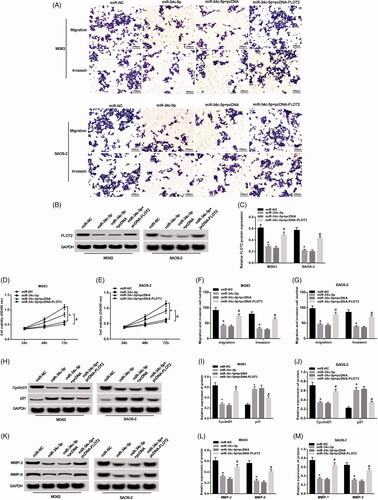
Overexpression of FLOT2 reverses the effect of miR-34c-5p overexpression on proliferation, migration and invasion of osteosarcoma cells
Western Blot results () showed that the expression levels of FLOT2, CyclinD1, MMP-2 and MMP-9 protein was significantly increased in MG63 and SAOS-2 cells of the miR-34c-5p + pcDNA-FLOT2 group compared to the miR-34c-5p + pcDNA group and the expression level of p21 protein was significantly decreased (p < .05). The MTT assay results () showed that the activity of osteosarcoma cells in the miR-34c-5p + pcDNA-FLOT2 group was significantly higher than that in the miR-34c-5p + pcDNA group (p < .05). The results of the Transwell assay () showed that the migration and invasion of MG63 and SAOS-2 cells in the miR-34c-5p + pcDNA-FLOT2 group were significantly higher than those in the miR-34c-5p + pcDNA group (p < .05). It can be seen that FLOT2 overexpression reverses the inhibitory effect of miR-34c-5p overexpression on proliferation, migration and invasion of osteosarcoma cells.
Figure 6. FLOT2 overexpression reverses the effect of miR-34c-5p overexpression on proliferation, migration and invasion of osteosarcoma cells. (A) The migration and invasion of osteosarcoma cells. (B, C) Relative expression of FLOT2 protein. (D, E) FLOT2 overexpression reverses the effect of miR-34c-5p overexpression on proliferation of MG63 and SAOS-2 cells. (F, G) FLOT2 overexpression reverses the effect of miR-34c-5p overexpression on migration and invasion of MG63 and SAOS-2 cells. (H–J) FLOT2 overexpression reverses the effect of miR-34c-5p overexpression on the expression of proliferation-related proteins in MG63 and SAOS-2 cells. (K–M) FLOT2 overexpression reverses the effect of miR-34c-5p overexpression on the expression of migration-invasive proteins in MG63 and SAOS-2 cells. Note: Compared with miR-NC group,*p < .05; Compared with miR-34c-5p + pcDNA group, #p < .05
Discussion
Osteosarcoma seriously affects the quality of life of patients. Early detection, early diagnosis and early treatment are the key factors [Citation11]. miRNA regulates the important pathophysiological processes in the development, progression, metastasis and chemotherapy of osteosarcoma, providing new ideas for targeted therapy of osteosarcoma [Citation12]. miR-34a-5p [Citation13] and miR-20a-5p [Citation14] can increase the chemosensitivity of osteosarcoma cells; miR-34a inhibits tumor invasion and metastasis of osteosarcoma by affecting C-IAP2 and Bcl-2 [Citation15]. In addition, miR-34a can also reduce the self-renewal ability, tumorigenic activity and invasive ability of cancer stem cells [Citation16]. miRNA-218 inhibits osteosarcoma cell migration and invasion by down-regulating TIAM1, MMP2 and MMP9 [Citation17]. miR-30a-5p [Citation18] and miR-33b [Citation19] are down-regulated in osteosarcoma and up-regulation of its expression inhibits osteosarcoma cell proliferation, invasion and promotes apoptosis. While miR-99a [Citation20]and miR-21 [Citation21] are highly expressed in osteosarcoma tissues and cells, down-regulation of their expression inhibits osteosarcoma cell proliferation, invasion and metastasis, and promotes apoptosis. Interestingly, miR-34c-5p functions as a carcinogen in various cancers, such as colon cancer [Citation22] and bladder cancer [Citation23]. However, increasing evidence indicates that miR-34c-5p acts as a tumor suppressor in various tumors, such as acute myeloid leukemia [Citation24]. miRNAs have different roles in different cancers, which are related to the tumor microenvironment. The role of miR-34c-5p in osteosarcoma has not been investigated. In the present study, the results showed that the expression of miR-34c-5p was significantly down-regulated in osteosarcoma tissues and cells. Overexpression of miR-34c-5p inhibited the expression of Cyclin D1, MMP-2 and MMP-9 proteins and inhibited the proliferation, migration and invasion of osteosarcoma cells. FLOT2 is widely distributed in organisms, involved in tumor progression, endocytosis and interaction of various protein molecules. FLOT2 inhibits cell proliferation, migration and invasion of gastric cancer cells [Citation25]. FLOT2 is up-regulated in gastric cancer and miR-449a reduces cell invasion by inhibiting the expression of Flot2 via TGF-β-mediated EMT [Citation26]. FLOT2 is highly expressed in oral squamous cell carcinoma [Citation27], nasopharyngeal carcinoma [Citation28] and non-small cell lung cancer [Citation29]. Knockdown of FLOT2 inhibits proliferation, migration and invasion of renal cell carcinoma cells [Citation30]. Overexpression of Flot2 promotes the growth and metastasis of hepatocellular carcinoma by regulating cell cycle and inducing epithelial-mesenchymal transition [Citation31]. Knockdown of Flot-2 inhibits the growth and invasion of human esophageal squamous cell carcinoma [Citation32]. Targeting FLOT2 via the Akt signaling pathway, miR-133 and miR-485 inhibits lung metastasis [Citation33,Citation34]. miR-34a inhibits melanoma cell proliferation and metastasis by directly targeting the FLOT2 gene [Citation35]. At present, there is no relevant research on FLOT2 in osteosarcoma. The results of this study showed that the expression level of FLOT2 was significantly upregulated in human osteosarcoma tissues and cells. Moreover, the proliferation, migration and invasion of osteosarcoma cell could be inhibited when FLOT2 expression was decreased. For the targeted regulation of miR-34c-5p and FLOT2, this study found that miR-34c-5p negatively targeted FLOT2. Overexpression of FLOT2 reversed the inhibitory effect of miR-34c-5p overexpression on proliferation, migration and invasion of osteosarcoma cells.
In summary, miR-34c-5p inhibited the proliferation, migration and invasion of osteosarcoma cells and its mechanism might be related to the targeted down-regulation of FLOT2, which will provide a new target for the diagnosis and treatment of osteosarcoma.
Disclosure statement
The authors declare that there are no conflict of interests.
References
- Li S, Sun W, Wang H, et al. Research progress on the multidrug resistance mechanisms of osteosarcoma chemotherapy and reversal. Tumour Biol. 2015;36:1329–1338.
- Liang W, Gao B, Fu P, et al. The miRNAs in the pathgenesis of osteosarcoma. Front Biosci. 2013;18:788–794.
- Wu Y, Ni Z, Yan X, et al. Targeting the MIR34C-5p-ATG4B-autophagy axis enhances the sensitivity of cervical cancer cells to pirarubicin. Autophagy. 2016;12:1105–1117.
- Wu H, Huang M, Lu M, et al. Regulation of microtubule-associated protein tau (MAPT) by miR-34c-5p determines the chemosensitivity of gastric cancer to paclitaxel. Cancer Chemother Pharmacol. 2013;71:1159–1171.
- Gu J, Wang G, Liu H, et al. SATB2 targeted by methylated miR-34c-5p suppresses proliferation and metastasis attenuating the epithelial-mesenchymal transition in colorectal cancer. Cell Prolif. 2018;51:e12455.
- Procopio M, Re M, Magliulo G, et al. Expression levels and clinical significance of miR-21-5p, miR-let-7a, and miR-34c-5p in laryngeal squamous cell carcinoma. BioMed Res Int. 2017;2017:3921258.
- Amaral AJ, Andrade J, Foxall RB, et al. miRNA profiling of human naive CD4 T cells links miR-34c-5p to cell activation and HIV replication. EMBO J. 2017;36:346–360.
- López JA, Alvarezsalas LM. Differential effects of miR-34c-3p and miR-34c-5p on SiHa cells proliferation apoptosis, migration and invasion. Biochem Biophys Res Commun. 2011;409:513–519.
- Feifei MA, Hao Y, Wang G, et al. The effect of up-regulating miR-34c-5p on the proliferation and invasion of nasopharyngeal carcinoma cell line HONE-1. Tianjin Med J. 2015;21(23):5378–5385.
- Fei W, Wang HB. Research progress of the relationship between Flot2 and malignant tumor. J Modern Oncol. 2017;25:816–819.
- Jones KB, Salah Z, Del MS, et al. miRNA signatures associate with pathogenesis and progression of osteosarcoma. Cancer Res. 2012;72:1865–1877.
- Kobayashi E, Hornicek FJ, Duan Z. MicroRNA involvement in osteosarcoma. Sarcoma. 2012;2012:359739.
- Pu Y, Zhao F, Li Y, et al. The miR-34a-5p promotes the multi-chemoresistance of osteosarcoma via repression of the AGTR1 gene. BMC Cancer. 2017;17:45.
- Zhao F, Pu Y, Cui M, et al. MiR-20a-5p represses the multi-drug resistance of osteosarcoma by targeting the SDC2 gene. Cancer Cell Int. 2017;17:100.
- Wen J, Zhao YK, Liu Y, et al. MicroRNA-34a inhibits tumor invasion and metastasis in osteosarcoma partly by effecting C-IAP2 and Bcl-2. Tumour Biol. 2017;39:1010428317705761.
- Zou Y, Huang Y, Yang J, et al. miR-34a is downregulated in human osteosarcoma stem-like cells and promotes invasion, tumorigenic ability and self-renewal capacity. Mol Med Rep. 2017;15:1631–1637.
- Jin J, Cai L, Liu ZM, et al. miRNA-218 inhibits osteosarcoma cell migration and invasion by down-regulating of TIAM1, MMP2 and MMP9. Asian Pac J Cancer Prev. 2013;14:3681–3684.
- Tao J, Cong H, Wang H, et al. MiR-30a-5p inhibits osteosarcoma cell proliferation and migration by targeting FOXD1. Biochem Biophys Res Commun. 2018;503:1092–1097.
- Zhou Y, Yang C, Wang K, et al. MicroRNA-33b inhibits the proliferation and migration of osteosarcoma cells via targeting hypoxia-inducible factor-1α. Oncol Res. 2017;25:397–405.
- Xie Y, Sun W, Xiao L, et al. Effect of MiR-99a inhibitor on the biological behavior of human osteosarcoma cells. Med J Wuhan Univ. 2018;39:36–41.
- Li C, Xu B, Miu X, et al. Inhibition of miRNA-21 attenuates the proliferation and metastasis of human osteosarcoma by upregulating PTEN. Exp Ther Med. 2018;15:1036–1040.
- Li N, Mao D, Cao Y, et al. Downregulation of SIRT6 by miR-34c-5p is associated with poor prognosis and promotes colon cancer proliferation through inhibiting apoptosis via the JAK2/STAT3 signaling pathway. Int J Oncol. 2018;52(5):1515–1527.
- Xu Z, Huang B, Zhang Q, et al. NOTCH1 regulates the proliferation and migration of bladder cancer cells by cooperating with long non-coding RNA HCG18 and microRNA-34c-5p. J Cell Biochem. 2019;120:6596–6604.
- Peng D, Wang H, Li L, et al. miR-34c-5p promotes eradication of acute myeloid leukemia stem cells by inducing senescence through selective RAB27B targeting to inhibit exosome shedding. Leukemia. 2018;32:1180–1188.
- Cao K, Xie D, Cao P, et al. SiRNA-mediated flotillin-2 (Flot2) downregulation inhibits cell proliferation, migration, and invasion in gastric carcinoma cells. Oncol Res. 2014;21:271–279.
- Li Q, Peng J, Li X, et al. miR-449a targets Flot2 and inhibits gastric cancer invasion by inhibiting TGF-β-mediated EMT. Diagn Pathol. 2015;10:202.
- Wen Q, Alnemah MM, Luo J, et al. FLOT-2 is an independent prognostic marker in oral squamous cell carcinoma. Int J Clin Exp Pathol. 2015;8:8236–8243.
- Zhao L, Lin L, Pan C, et al. Flotillin-2 promotes nasopharyngeal carcinoma metastasis and is necessary for the epithelial-mesenchymal transition induced by transforming growth factor-β. Oncotarget. 2015;6:9781–9793.
- Wen Q, Wang W, Chu S, et al. Flot-2 expression correlates with EGFR levels and poor prognosis in surgically resected non-small cell lung cancer. PLoS One. 2015;10:e0132190.
- Yan Y, Yang FQ, Zhang HM, et al. Up-regulation of flotillin-2 is associated with renal cell carcinoma progression. Tumour Biol. 2014;35:10479–10486.
- Wang CH, Zhu XD, Ma DN, et al. Flot2 promotes tumor growth and metastasis through modulating cell cycle and inducing epithelial-mesenchymal transition of hepatocellular carcinoma. Am J Cancer Res. 2017;7:1068–1083.
- Li L, Peng X, Wang Y, et al. High expression of Flot-2 promotes the development and invasion of esophageal squamous cell carcinoma. Chinese J Histochem Cytochem. 2017;2:66–70.
- Wei G, Xu Y, Peng T, et al. miR-133 involves in lung adenocarcinoma cell metastasis by targeting FLOT2. Artif Cells Nanomed Biotechnol. 2018;46:224–230.
- Mou X, Liu S. MiR-485 inhibits metastasis and EMT of lung adenocarcinoma by targeting Flot2. Biochem Biophys Res Commun. 2016;477:521–526.
- Liu R, Xie H, Luo C, et al. Identification of FLOT2 as a novel target for microRNA-34a in melanoma. J Cancer Res Clin Oncol. 2015;141:993–1006.