
 ?Mathematical formulae have been encoded as MathML and are displayed in this HTML version using MathJax in order to improve their display. Uncheck the box to turn MathJax off. This feature requires Javascript. Click on a formula to zoom.
Abstract
Effective and sustained release formulations of antimetabolite 5-Flurouracil (5Fu) for ocular fibrosis are highly desirable for success of glaucoma filtration surgery (GFS). However, burst release and rapid clearance of carriers and/or drug still exist as major limitations. Here, we report the feasibility of encapsulating 5Fu in sustained release carrier of poly (d,l-lactide-co-glycolide) (PLGA) microspheres (MPs) and impregnating in a hyaluronic acid (HA) hydrogel as unit dose formulation. In order to optimize the encapsulation process by solid-in-oil-in-water emulsion technique, the effects of PLGA end groups, PLGA concentration, and 5Fu loading were studied systematically. The MPs were extensively characterized for surface morphology, particle size distribution, thermal properties, crystallinity, residual solvent, syringeability, and in vitro release. The optimized formulation of MPs was ~94 μm in size and released 5Fu in a sustained manner for up to two weeks. In our concept, MPs are dispersed first in HA solution and then cross-linked in situ after injection into the conjunctival space. Thus, syringeability experiments were also carried out with 5Fu-loaded MPs and 0.5% HA solution. To further prolong 5Fu release, a derivatized HA with methacrylic anhydride was photocross-linked along with 5Fu-loaded PLGA MPs to develop the composite hydrogel formulation. Our experimental results show that 5Fu release can be effectively controlled by PLGA MPs-loaded HA composite hydrogel for longer therapeutic benefits. These composite hydrogel systems improve the ocular residence time of the carrier and the drug, and hence could be potentially explored as injectable unit dose formulation for GFS.
Public Interest Statement
Glaucoma is a common eye disease that leads to blindness and affects large number of population worldwide. The surgical treatment often fails due to several limitations of the drug, 5-Fluorouracil (5Fu), that is injected into the eye either during or after glaucoma surgery. In our work, we have addressed these issues by developing a potential delivery system for 5Fu via combination of two different carriers. Micron-sized particles are first loaded with 5Fu and then entrapped inside a gel network. This combined system is nontoxic and gets safely disintegrated inside body after treatment. It was investigated in detail for its material properties and our study shows that 5Fu is released slowly from particle–gel combined system within the desired therapeutic range for almost two weeks. This would ensure that patients do not need multiple injections. Moreover, our system, upon injection inside eye, will provide localized treatment avoiding any side effects.
Competing interests
The authors declare no competing interest.
1. Introduction
Glaucoma is one of the most common ocular diseases leading to irreversible vision loss and is characterized by high fluid pressure inside the eye resulting in optical nerve damage (Gooch et al., Citation2012). Glaucoma is currently the second leading cause of blindness worldwide (Kingman, Citation2004). In the year 2012, it was estimated that around 60 million people were affected with glaucoma globally and an estimated 8.4 million of them were blind as a result of it. This number is predicted to increase by approximately 33% to 80 million affected patients by the year 2020 (Cook & Foster, Citation2012; Quigley, Citation2011; Quigley & Borman, Citation2006).
Current clinical practice involves reduction of intraocular pressure (IOP) by medications along with surgical approaches. One of the conventional surgical methods is glaucoma filtration surgery (GFS) that introduces an alternative path for drainage of excess fluid thus reducing IOP. A small opening is created through the sclera in the drainage angle of the eye and this surgically created channel provides an alternative path for drainage of aqueous humor outside the eye, thus bypassing the clogged or blocked channels of the trabecular meshwork (Green, Wilkins, Bunce, & Wormald, Citation2014; Wormald, Wilkins, & Bunce, Citation2001).
However, after the surgery, the body’s natural healing response may lead to the blockage of the surgically created channel, as subconjunctival fibroblasts start to proliferate. The resulting fibrosis leads to the blockage and to failing and encysted blebs. In this healing process, proliferation of fibroblasts from the episclera and Tenon’s capsule plays an important role. To prevent the scarring of filtering blebs, subconjunctival injections of 5Fu, an antimetabolite, are given either post-operatively or as a single intraoperative injection (How et al., Citation2010; Mead, Wong, Cordeiro, Anderson, & Khaw, Citation2003).
5Fu is a fluorinated pyrimidine analog, mainly used as an anticancer drug and functions by interfering with nucleoside metabolism, inhibiting thymidylate synthase (Jacob, LaCour, & Burgoyne, Citation2001; Longley, Harkin, & Johnston, Citation2003; Mahoney, Raghunand, Baggett, & Gillies, Citation2003). Recently, 5Fu was widely reported as an effective antimetabolite in clinical applications of ophthalmology to inhibit proliferation of fibroblasts.
The current mode of use of 5Fu has several limitations (Chiang, Tung, Lu, & Yeh, Citation2001; Cui, Sun, LI, Huang, & Yang, Citation2008; Huhtala et al., Citation2009; Lee, Hersh, Kersten, & Melamed, Citation1987; Peyman, Conway, Khoobehi, & Soike, Citation1992). It is not selective toward fibroblast inhibition, and indeed affects other healthy ocular cells. Also, the drug is not retained for more than a few hours at the site of administration, thus necessitating multiple injections, to which patients do not comply well. Moreover, the high dose administered locally results in ocular surface complications.
Clearly, there is a need to develop a local delivery system for 5Fu that would sustain its release at target site for prolonged periods of time and reduce multiple dosing, in addition to lowering toxicity. Several polymeric drug delivery microcarriers have been investigated to achieve these targets (Arias, Citation2008; Kimura & Ogura, Citation2001; Rocíoh, Citation2011; Zimmer & Kreuter, Citation1995). Among them, poly (d,l-lactide-co-glycolide) (PLGA)-based biodegradable microspheres MPs are advantageous due to numerous therapeutic benefits. PLGA is a copolymer of poly lactic acid (PLA) and poly glycolic acid (PGA) and its biodegradability, nontoxicity, and tunable mechanical and erosion properties have led to its extensive usage as drug delivery carriers (Chang et al., Citation2011; Choi, Seo, & Yoo, Citation2012; Makadia & Siegel, Citation2011; Mundargi, Babu, Rangaswamy, Patel, & Aminabhavi, Citation2008).
Several researchers have reported improved outcomes in GFS using PLGA carriers loaded with 5Fu as an antiscarring agent. Lu, Chang, Chiang, Yeh, and Chou (Citation2000) prepared 5Fu-loaded PLGA MPs by an o/o emulsification method to study the effects of subconjunctival retention of these MPs inside rabbit eyes. It was found that improvement in the success of GFS using 5Fu-loaded PLGA MPs was dose dependent. Yeh and co-workers investigated the formulation parameters for preparing 5Fu-encapsulated PLGA MPs and characterized them in vitro and in vivo to evaluate their potential for ocular administration in GFS (Chiang et al., Citation2001; Yeh, Tung, Lu, Chen, & Chiang, Citation2001). The MPs were prepared by o/o emulsion or solvent extraction method. The MPs had a high entrapment efficiency of 76% with practical loading of 10%. After a high initial burst, 5Fu was released over 21 days in a sustained manner with first-order release kinetics. In another study, 5Fu-loaded PLA MPs disks were developed by Cui et al. (Citation2008) and evaluated for sustained drug release inside rabbit eyes for GFS. Drug-loaded PLA MPs were accumulated by repeating cycles of addition and precipitation using petroleum benzine, and then gently compressed to form disks. After initial burst release on day 1, the release rate was markedly slowed down from day 2 onwards, with slow release sustained over 90 days.
These earlier reports demonstrated the potential of obtaining sustained release of 5Fu using biodegradable PLGA MPs. However, the critical problems associated with initial high burst release and rapid clearance were not solved. High burst release would not only cause local toxicity but also lead to drug levels below the therapeutic index in the later stages of release period. Rapid clearance of drug and microcarriers (following the high initial burst) leads to the need for frequent administration or repeating doctor visits for the patient. These drawbacks can be overcome using composite hydrogel systems, wherein drug-loaded microcarriers are homogeneously dispersed within a hydrogel matrix. Composite hydrogels have been shown to enable sustained release of drug for prolonged periods by reducing burst release and providing an additional barrier to the diffusion of drugs (Bae, Go, Park, & Lee, Citation2006; Caicco et al., Citation2013; Hou et al., Citation2008; Joung, Choi, Park, & Park, Citation2007; Lagarce et al., Citation2005; Lin, Sun, Jiang, Zan, & Ding, Citation2007; Nath, Linh, Sadiasa, & Lee, Citation2014; Ranganath, Kee, Krantz, Chow, & Wang, Citation2009; Zhao, Guo, & Ma, Citation2014). The aforementioned combination strategy is advantageous over MPs or gels alone because it improves drug’s half-life, preserves their bioactivity, improves the stability of MPs, and enhances the bioavailability of drugs by preventing the migration of drug-loaded carriers away from the target site (Hoare & Kohane, Citation2008; Mufamadi et al., Citation2011). In our previous work, we have shown that hyaluronic acid (HA)-based composite hydrogel systems can be used as sustained release carriers for ocular drug delivery applications (Widjaja et al., Citation2014). HA is a natural polymer composed of repeating disaccharide units of β-d-glucuronic acid (GlcUA) and N-acetyl-β-d-glucosamine (GlcNAc) linked by β (1–3) and β (1–4) glycosidic bonds and belongs to the class of glycosaminoglycans or mucopolysaccharides. In the human body, HA is present in the vitreous humor (eye’s posterior cavity) as well as in other locations, and plays a significant role in many biological functions on account of its nontoxicity, viscoelasticity, and mucoadhesive properties (Jin, Ubonvan, & Kim, Citation2010; Kogan, Soltes, Stern, & Gemeiner, Citation2007).
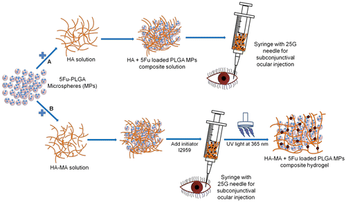
Composite systems of PLGA MPs with HA hydrogels have recently been explored mainly for tissue bulking and engineering applications, whereas drug delivery applications have not yet been investigated (Galeska et al., Citation2005; Ju, Wen, Gou, Wang, & Xu, Citation2014; Patterson, Stayton, & Li, Citation2009; Tous et al., Citation2012; Wang et al., Citation2011; You et al., Citation2014). In the present work, we have developed a composite system of 5Fu-loaded PLGA MPs entrapped within HA hydrogel as an ocular drug delivery system for post-GFS applications. The primary objective of our study was to provide sustained release of 5Fu from PLGA MPs impregnated inside the HA hydrogel network; the composite is expected to increase the ocular residence time of drug and thus its bioavailability for longer durations, consistent with the period of fibrosis formation. We have optimized the encapsulation of 5Fu into PLGA MPs by s/o/w emulsion and solvent evaporation technique. The effects of process parameters viz., PLGA end group, concentration, and 5Fu theoretical loading, on the yield, encapsulation efficiency (EE), practical loading, and in vitro release profile of PLGA MPs were evaluated. MPs with highest practical loading of 5Fu were selected for incorporation into HA solution as well as HA hydrogel and investigated for in vitro release study. The feasibility of composite system of 5Fu-loaded PLGA MPs in HA solution for intraocular injections was also evaluated via multiple syringeability experiments. Schematic in shows the development of proposed PLGA MPs and HA composite formulation for ocular delivery. The optimized 5Fu-loaded PLGA MPs were infused in HA solution and hydrogel for prolonging local residence time at target site.
Figure 1. Schematic showing the development and ocular delivery of composite HA formulation.
Notes: (A) 5Fu-loaded PLGA MPs infused in HA solution can be administered using 25G needle and (B) 5Fu-loaded PLGA MPs infused in HA-MA solution can be administered followed by UV cross-linking to form MPs-impregnated hydrogel matrix system.
2. Materials
HA (sodium salt, Mw = 1 × 10⁶ g mol−1) was purchased from Samich (HK) Limited, China. Chemicals methacrylic anhydride (MA), poly (vinyl alcohol) (PVA) (87–90% hydrolyzed, average MW = 30,000–70,000 g mol−1), and 5Fu (MW = 130.1 g mol−1) were obtained from Sigma-Aldrich, Singapore. PLGA, Purasorb PDLG 5004A, and Purasorb PDLG 5004E (L:G = 50:50, 0.4A PLGA is acid terminated and 0.4E PLGA is ester terminated, with inherent viscosity, IV, = 0.4 dl/g PLGA) were purchased from PURAC Asia Pacific Pte Ltd, Singapore. Chemicals sodium chloride, sodium phosphate, potassium phosphate monobasic anhydrous, potassium chloride, sodium hydroxide, hydrochloric acid, mannitol, and deuterated chloroform were obtained from Sigma-Aldrich, Singapore and were used directly without purification. 2-methyl-1-[4-(hydroxyethoxy) phenyl]-2-methyl-1-propanone or Irgacure 2959 (I2959) was purchased from Ciba Speciality Chemicals (Singapore) Pte Ltd, Singapore. Dichloromethane (DCM) and methanol were obtained from Fisher Scientific, Singapore. Sieves (106 μm and 150 μm) and sonication probe (3 mm) were obtained from Cole Parmer, USA. Syringe filters (0.22 μm) were purchased from Merck Pte Ltd (Merck Millipore Division), Singapore.
3. Methods
3.1. Preparation of 5Fu-loaded PLGA microparticles
5Fu-loaded PLGA MPs were prepared using modified solvent evaporation method of s/o/w single emulsion technique (Faisant, Siepmann, & Benoit, Citation2002). Commercial 5Fu powder was crushed using a mortar and pastel to reduce its grain size and was dispersed in DCM. log P of 5Fu is reported to be around −0.89 (Naguib, Kumar, & Cui, Citation2014; Nasr, Ghorab, & Abdelazem, Citation2015). The mixture was sonicated (3 mm Probe, SONICS Vibra cell, USA) for 2 min at 30% amplitude to further reduce the crystals of 5Fu and dispersed them homogeneously in DCM. PLGA (0.4A IV or 0.4E IV), 20% w/v, was added into this suspension and vortexed vigorously to completely dissolve the PLGA. The suspension was then sonicated for 2 min and immediately added into the continuous aqueous phase of PVA (40 ml, 1% w/v, pH adjusted to 1.7 using 5 M HCl) using a glass pasteur pipette to form the emulsion. 5Fu is a weak acid and it pKa is reported to be around 7.6–8 (Kondo & Aroie, Citation1989; Mahoney et al., Citation2003). PVA was dissolved in DI water at 85°C, filtered through 0.22-μm filter, and pH was adjusted to 1.7. The emulsion was stirred at 1,000 rpm, room temperature (RT) to evaporate the solvents for 1.5 h. After 1.5 h, 60 ml of PVA (1% w/v, pH adjusted to 1.7) was added into the emulsion and evaporation was further continued for 3.5 h. The drug-loaded MPs were collected using centrifuge (Thermo Electron Corporation B4i Multifunctional, USA) at 8,000 rpm (rcf 7, 969×g) for 6 min at RT. The MPs were resuspended using 10 ml of ultrapure water and washed twice. The MPs were finally suspended in mannitol (1% w/v in ultrapure water) and lyophilized for 24 h.
Different batches of 5Fu-loaded PLGA MPs were prepared to understand the effect of PLGA end group, theoretical loading of 5Fu, and PLGA concentration as summarized in . All the MPs formulations were stored in dry cabinet at 22°C until further characterization. From these batches, the batch with highest practical loading of 5Fu (corresponding to lowest amount of PLGA MPs containing 1 mg 5Fu) with desirable size range (<100 μm) was selected for further studies with HA solution and hydrogel.
Table 1. Formulation parameters of 5Fu-loaded PLGA MPs
3.2. Synthesis of HA hydrogels
HA was first modified into its methacrylated derivative, HA-MA, using MA as described previously (Nettles, Vail, Morgan, Grinstaff, & Setton, Citation2004; Smeds et al., Citation2000). HA powder was dissolved in DI water to prepare 1.5% (w/v) solution and pH was adjusted to 8 using 5 M NaOH while slowly adding 20 M excess of MA to it. The reaction was allowed to continue for 2 h at RT while maintaining the pH at 8 and then stored at 4°C for 24 h. HA-MA solution was dialyzed against 0.1 M NaCl solution using a dialysis membrane (Spectra/Por 6 dialysis tubing, 10 kDa molecular weight cut-off (MWCO), 29 mm diameter, flat width 45 mm) for 48 h followed by 2 cycles of dialysis against alternating solutions of 1:4 C2H5OH– H2O (v/v) and ultrapure H2O for 24 h before lyophilization for 72 h (Christ Alpha 1–4 LSC, Germany). After freeze drying, 4% (w/v) solution of HA-MA was prepared in PBS, pH 7.4 and 0.4% (w/w) of photoinitiator I2959 was added into it. This macromer solution with initiator was then exposed to UV lamp (VL-8.LC, 1 × 8 W, Vilber Lourmat, France) at 365 nm for 10 min (light intensity 720 μW/cm2 at 15 cm) for cross-linking to develop HA-MA hydrogels.
3.3. Preparation of composite hydrogels
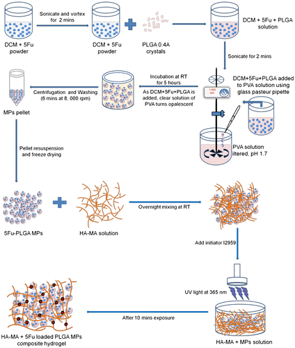
Modified HA powder, HA-MA, was dissolved in PBS buffer to prepare 4% (w/v) solution and allowed to stir overnight at RT. Next day, 25 mg of 5Fu-loaded PLGA MPs (containing 1 mg/ml 5Fu) were weighed and mixed with HA-MA solution (polymer to particles mass ratio of 1.6:1). Upon mixing, HA-MA + 5Fu-PLGA MPs solution was cross-linked using UV light as described above. Schematic in shows the fabrication of 5Fu-loaded PLGA MPs, incorporation in HA-MA solution, and subsequent cross-linking by UV light to obtain composite hydrogel system.
Figure 2. Schematic showing the fabrication of 5Fu-loaded PLGA MPs.
Notes: The MPs were prepared by via s/o/w emulsion technique by varying PLGA end groups, PLGA concentration, and drug loading to achieve better EE. 5Fu-encapsulated MPs were infused in photocross-linked HA hydrogel.
3.4. Characterization of 5Fu-loaded PLGA MPs
3.4.1. Yield
After freeze drying, 5Fu-loaded MPs were sieved using 150-μm and 106-μm sieves to remove polymeric debris and small fibers. The dry mass of MPs was recorded before and after sieving to estimate the % yield. The % yield of 5Fu-loaded MPs was calculated from the freeze-dried and sieved MPs using the following equation:
3.4.2. Morphology and size distribution
The shape and surface morphology of 5Fu-loaded PLGA MPs were observed under SEM (JSM 6360A Jeol, Japan). Dry MPs samples were sputter coated with gold for 80 s and then imaged under different magnifications at an acceleration voltage of 5–10 kV. For size distribution, dry MPs samples were suspended in Tween 80 (0.25% v/v in DI water), ultrasonicated, and run in Fritsch Laser Particle Analyzer (Analysette 22 MicroTec plus, Fritsch Asia Pacific Pte Ltd, Singapore) for 5 measurements with 100 scans each. The size distribution of MPs was obtained with respect to % volume of sample and volume mean and mode diameters were reported.
3.4.3. Determination of loading and EE
Dry-sieved 5Fu-loaded PLGA MPs were dissolved in DCM and PBS was added into it in a separating funnel. The contents of the funnel were vigorously shaken for few minutes to mix the PBS solution and DCM. The solutions were then allowed to settle down at RT for 5–10 min. The immiscible phases of PLGA-DCM and 5Fu-PBS separate. This vigorous shaking and phase separation were continued for 2 more cycles. After the 3rd cycle, the top phase of 5Fu-PBS was collected separately in a tube. To the bottom phase of PLGA-DCM, fresh PBS was added. These above steps were repeated for 5 cycles. From the 5Fu-PBS solution, collected from 5 cycles, 1-ml aliquot was withdrawn and analyzed using HPLC system (Agilent 1200 series, Agilent Technologies Singapore Pte Ltd).
A reverse phase Eclipse-XDB C18 column (5 μm, 4.6 mm ID × 250 mm) was used with the mobile phase of H2O:CH3OH at 90:10 (v/v) ratio. The column was maintained at a temperature of 25°C. Each sample was run at a flow rate of 1.0 ml/min for 5 min including 1 min of post-run with the detector wavelength set at λ = 266 nm. The retention time of 5Fu was ~3.3 min. Collected data were analyzed using Agilent’s offline Chemstation software. 5Fu solutions with known concentrations (0.025–25 μg/ml) were prepared by dissolving the crushed 5Fu powder in PBS buffer to obtain a standard curve (R2 = 0.999). Concentrations of 5Fu for the test samples were calculated using this calibration of HPLC system. These data from HPLC were used to estimate the amount of 5Fu entrapped inside PLGA MPs and to calculate the practical loading and % EE using the following equations:
3.4.4. Differential scanning calorimetry
The thermal properties of free 5Fu powder and 5Fu-loaded inside PLGA MPs were evaluated using Differential scanning calorimetry (DSC) (Q10 V9.9 Build 303, TA Research Instruments, UK). Each sample was taken in a standard aluminum pan, crimped with the lid, and run for 2 ramp cycles. In every cycle, samples were cooled at 10°C/min from RT to 0°C, equilibrated at this temperature for 2 min, heated at 2°C/min to 330°C, equilibrated at this temperature for 2 min, and cooled back to 0°C at 10°C/min. An empty pan, sealed in aluminum pan similar to samples, was used as the reference. TA’s Universal analysis software was used to analyze the melting point of 5Fu (T m) and glass transition temperature of PLGA (T g) from the DSC thermograms.
3.4.5. X-ray diffraction
The crystalline nature of 5Fu, before and after encapsulation inside PLGA MPs, was evaluated from X-ray Diffraction (XRD) spectra obtained on a powder X-ray diffractometer (LabX 6,000, Shimadzu (Asia Pacific) Pte Ltd, Singapore) with Cu–Kα radiation at λ = 1.5406 Å. Scans for each sample were recorded in continuous mode with 2θ ranging from 10° to 50°, at a step size of 0.02 2θ.
3.4.6. Nuclear magnetic resonance (NMR)
Proton (1H) NMR spectrum for 5Fu-loaded PLGA MPs was obtained using Bruker NMR Spectrometer (Bruker DRX 400, Germany) at 400 MHz to determine the presence of residual DCM in dried PLGA MPs. The two samples of MPs tested were obtained after vacuum oven drying at 40°C for either 2 or 4 days post-24 h of freeze drying. NMR samples were prepared by dissolving the PLGA MPs in deuterated chloroform inside NMR tubes and analyzed for 1H spectral data with 200 scans per sample.
3.4.7. Syringeability
HA solutions with varying concentrations (0.1, 0.25, 0.5, 1, and 2.3%) were prepared in PBS, pH 7.4, and centrifuged (Mikro 20, Hettich, Germany) to remove any air bubbles. 0.5 ml of each solution was injected out into a fresh vial and mixed with ~25 mg of 5Fu-loaded PLGA MPs (5-mg MPs per 100-μl HA) to form composite solution. Syringe (1 ml) was then used to collect the composite solution of HA and MPs without creating any air bubbles. The MPs-loaded syringe was attached to hypodermic needles of varying gages (23G, 25G, and 27G) and composite solution was injected out carefully in 100-μl volumes and weighed. The mass of composite solution injected out per 100 μl was recorded and 5Fu, extracted from the entrapped MPs, was analyzed using HPLC.
3.4.8. In vitro drug release study
5Fu release studies were conducted with PLGA MPs alone and PLGA MPs incorporated in HA solution as well as HA-MA hydrogels to evaluate the efficacy of each formulation in sustaining the release within therapeutic index. Dry PLGA MPs (containing 1-mg 5Fu) were weighed and placed directly into a CE dialysis membrane (10 kDa MWCO, 2.9 cm diameter, flat width 4.5 cm), immersed in PBS, and incubated inside an orbital shaker incubator (LM-570RD, Yihder Technology, Taiwan) at 50 rpm maintained at 37°C.
For incorporation into HA solutions and HA-MA hydrogels, the 20% PLGA concentration (0.4A) with 20% 5Fu theoretical loading formulation was chosen for in vitro release studies based upon its practical loading, yield, and size. For HA solution, HA powder was dissolved in PBS (0.5% w/v) and mixed manually with 5Fu-loaded PLGA MPs (containing 1-mg 5Fu). For hydrogels, HA-MA powder was dissolved in PBS (4% w/v), mixed manually with 5Fu-loaded PLGA MPs (containing 1-mg 5Fu), and cross-linked under UV light at 365 nm for 10 min in the presence of I2959. These composite solutions and hydrogels were transferred into dialysis membrane, immersed in PBS, and incubated at 37°C inside shaker incubator.
One milliliter aliquots were withdrawn at fixed time intervals from the release medium of the samples, filtered through 0.2-μm syringe filter, and analyzed by HPLC using the calibration as discussed earlier. At every time point, the release medium was completely replaced with fresh PBS buffer.
3.4.9. Statistical analysis
Comparison between two samples for significant differences was derived from statistical analysis using Student’s two-tailed t-test. Differences were considered to be statistically different at p < 0.05.
4. Results and discussion
4.1. Effect of PLGA end group
PLGA chain ends can be either acid terminated (A) or ester terminated (E). In our study, we used 0.4A and 0.4E PLGA (L:G 50:50) MPs, with a theoretical 5Fu loading of 10%, and they were compared for in vitro release of 5Fu to understand the effect of end group type. shows the yield, EE, and amount of MPs containing 1-mg 5Fu for 0.4A and 0.4E PLGA MPs loaded with 5Fu. 5Fu-loaded PLGA MPs were extracted using the solvent phase separation method to estimate the practical loading and EE. The concentration of entrapped and unentrapped 5Fu was calculated using HPLC system. A standard curve (R2 = 0.999) was prepared using known concentrations of 5Fu in PBS buffer and the calibration was used to estimate the concentration of drug samples from MPs.
Table 2. Comparison of acid- (0.4A) and ester (0.4E)-terminated PLGA MPs with respect to yield, EE, and amount of MPs containing 1-mg 5Fu
As we can observe, % yield was almost similar in both the batches. % EE was slightly higher in 0.4E MPs and thus the amount of MPs containing 1-mg 5Fu was lesser in quantity. This might possibly be due to the relatively more hydrophilic nature of 0.4A MPs than 0.4E MPs that result in loss and lower entrapment of 5Fu crystals in the former during fabrication.
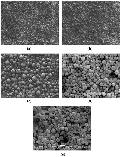
shows the SEM images of 5Fu-loaded PLGA MPs fabricated via s/o/w encapsulation technique with varying end groups. The MPs were spherical in shape and the surface was smooth. There was no distinct difference in the morphology of MPs between 0.4A and 0.4E.
Figure 3. SEM images of 5Fu-loaded MPs varying in PLGA chain end groups.
Notes: (a) acid-terminated 0.4A PLGA MPs and (b) ester-terminated 0.4E MPs.

To understand the release behavior of 5Fu from MPs prepared using different end groups, we performed in vitro release studies and data are displayed in . The % cumulative release of 5Fu from PLGA MPs, varying in end groups, indicates 5Fu release following a sigmoidal pattern, with a sharp curve in the case of 0.4A MPs, whereas an extended curve is seen in the case of 0.4E MPs. In other words, the onset of an enhanced release phase is seen on day 4 for the 0.4A PLGA, and on day 21 for the 0.4E PLGA MPs. The lactide-to-glycolide (L:G) ratio of PLGA MPs used in this work is 50:50; they absorb and get hydrated quickly as soon as they are exposed to the aqueous medium. The polymer chains in the matrix of PLGA MPs swell as the aqueous medium moves in and make the MPs porous leading to diffusion and release of 5Fu (Fredenberg, Wahlgren, Reslow, & Axelsson, Citation2011; Makadia & Siegel, Citation2011).
Figure 4. In vitro release profile of 5Fu from PLGA MPs showing faster release from acid-terminated end group as compared to ester-terminated MPs.

The observed release pattern is attributed to differences in the onset of erosion of PLGA due to variable end groups, with the acid-terminated chains of PLGA starting to erode faster than ester-terminated polymers because of the higher hydrophilicity or water-absorbing capacity and the acid-catalytic nature of the random chain scission process of bulk degradation of PLGA chains (Fredenberg et al., Citation2011; Samadi et al., Citation2013; Siegelr & Rathbonem, Citation2012). Hence, 5Fu was released in a faster rate from acid-terminated PLGA MPs, 100% of drug being released in 12 days from 0.4A MPs as compared to 33 days from 0.4E MPs.
4.2. Effect of PLGA concentration
The therapeutic daily dosage requirement for subconjunctival administration of 5Fu is reported to be 1 μg per day while the toxic concentration is 100 μg/ml (Wang, Tucker, Roberts, & Hirst, Citation1996). It was observed that the amount of 5Fu released per day from 0.4A PLGA MPs was above the therapeutic requirement for 75% of complete release period while 0.4E MPs released higher amounts only toward later stages (Figure S1). Hence, 0.4A PLGA MPs were selected for further studies investigating the effect of PLGA concentration and 5Fu loading.
To evaluate the effect of PLGA concentration, 0.4A PLGA MPs, with a theoretical drug loading of 10%, were prepared with 5, 10, 20, 30, and 40% PLGA and compared for in vitro drug release. shows the yield, EE, and amount of MPs containing 1-mg 5Fu for 0.4A PLGA MPs prepared with different concentrations of PLGA.
Table 3. Comparison of yield, EE, and amount of MPs containing 1-mg 5Fu for 0.4A PLGA MPs fabricated with varying PLGA concentrations
Our data, after varying the PLGA concentration, indicate that MPs prepared using 5% w/v and 10% w/v PLGA were not suitable to encapsulate 5Fu successfully. This was mainly because of the lower polymer viscosity that produced smaller emulsion droplets resulting in smaller size MPs. Since we incorporated 5Fu into MPs by physical encapsulation in its crystalline form via s/o/w single emulsion technique, hence, the MPs have to be relatively bigger than the size of 5Fu crystal grains for successful encapsulation.
For 30 and 40% PLGA concentrations, the yield using 150- and 106-μm sieves was very low due to the presence of MPs with size larger than 200 μm. Hence, % EE was less and the amount of PLGA MPs containing 1-mg 5Fu was high. shows the SEM images of 5Fu-loaded PLGA MPs with varying PLGA concentrations. The MPs were spherical in shape and the surface was smooth. There was no distinct difference in the morphology of MPs between different concentrations of PLGA. Strikingly, we can clearly observe the increase in size of MPs prepared using 5–40% PLGA concentrations.
Figure 5. SEM images of 5Fu-loaded 0.4A PLGA MPs prepared using different concentrations of PLGA.
Notes: (a) 5%, (b) 10%, (c) 20%, (d) 30%, and (e) 40%.
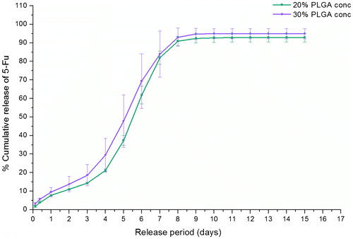
Further, to investigate the in vitro release profile of 5Fu-loaded MPs, we have selected MPs prepared using 20 and 30% PLGA concentrations due to better 5Fu encapsulation (see ). The MPs’ batches prepared using 5, 10, and 40% PLGA were not included in release studies due to low EE and yield. The release profiles () of MPs, prepared using 20 and 30% PLGA, indicated that ~100% of 5Fu was released by day 9 with no significant dependence of this rate on PLGA concentration. Our data suggest that 5Fu release rate was independent of the differences in practical drug loading (amount of 5Fu encapsulated inside 1 mg of MPs after fabrication) and MPs size distribution.
Figure 6. In vitro release profile of 5Fu from 0.4A PLGA MPs showing similar rate of release from 20 to 30% PLGA concentrations.
4.3. Effect of % 5Fu loading
To evaluate the effect of theoretical 5Fu loading, 0.4A PLGA MPs were prepared with 10, 15, and 20% 5Fu loading and compared for in vitro drug release. shows the yield, EE, and amount of MPs containing 1-mg 5Fu for 0.4A PLGA MPs loaded with different concentrations of 5Fu. As we can observe, the % yield and % EE of 10% 5Fu loading batch MPs was higher than that of 15 and 20% MPs. However, the amount of MPs containing 1-mg 5Fu was not lower than the other two batches.
Table 4. Comparison of yield, EE, and amount of MPs containing 1-mg 5Fu for 0.4A PLGA MPs fabricated with varying % theoretical loadings of 5Fu
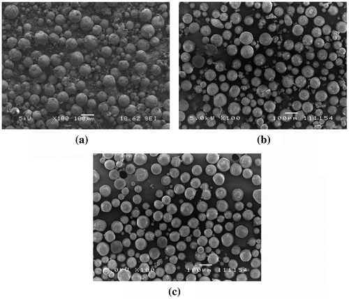
PLGA MPs with 20% theoretical loading have the highest practical loading. This indicates that there needs to be a balance between the amount of 5Fu used for encapsulation inside MPs and % EE to obtain higher levels of practical loading. shows the SEM images of 5Fu-loaded PLGA MPs with varying theoretical loadings of 5Fu. The MPs were spherical in shape and the surface was smooth. There was no distinct difference in the morphology of MPs between different theoretical loadings of 5Fu.
Figure 7. SEM images of 5Fu-loaded 0.4A PLGA MPs prepared using different theoretical loadings of 5Fu.
Note: (a) 10%, (b) 15%, and (c) 20%.
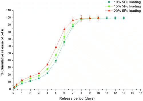
shows the % cumulative release of 5Fu from PLGA 0.4A with varying theoretical 5Fu loadings. As we can observe, the release trend was similar in all the samples. Further, higher the theoretical loading of 5Fu, faster was the release rate. The release rate differences were mainly observed between days 3 and 7 among batches B-I and B-If when the theoretical loading was doubled from 10 to 20%, respectively. This might be because of the lower % EE of batch B-1f than B-I.
Figure 8. In vitro release profile of 5Fu from 0.4A PLGA MPs showing increase in rate of release with increase in theoretical loading of 5Fu from 10 to 20%.
From our encapsulation and in vitro release data, 5Fu-loaded PLGA MPs, with EE of around 20% and in vitro release of almost 100% by day 8, were selected for further HA composite formulation studies and extensive characterizations involving particle size, thermal analysis, drug crystallinity by XRD, residual solvent analysis by NMR, and syringeability were performed. Our selected formulation released 1–29 μg of 5Fu per day maintaining the therapeutic dosage up to 13 days.
4.4. Size distribution
The size distribution of MPs with respect to % volume of sample was obtained using Fritsch Laser Particle Analyzer and the plot is shown in . The volume mean diameter was found to be 94.2 ± 0.2 μm, suitable for ocular injections for back of the eye applications. The PLGA MPs were very stable and exhibited a unimodal, narrow size distribution. We believe the uniform negative charge distribution and narrow size distribution, achieved on account of the fabrication parameters, are responsible for the stability of the particles.
Figure 9. Particle size analysis of 0.4A PLGA MPs (20% PLGA, 20% 5Fu loading) showing narrow distribution profile with an average diameter of ~94 μm.
Note: Q3(x) [%] represents the percent of complete sample volume that is filled with MPs smaller in size than “x” μm and dQ3(x) [%] indicates the percent of complete sample volume that is filled with MPs having a diameter between “x” and “y” μm.
![Figure 9. Particle size analysis of 0.4A PLGA MPs (20% PLGA, 20% 5Fu loading) showing narrow distribution profile with an average diameter of ~94 μm.Note: Q3(x) [%] represents the percent of complete sample volume that is filled with MPs smaller in size than “x” μm and dQ3(x) [%] indicates the percent of complete sample volume that is filled with MPs having a diameter between “x” and “y” μm.](/cms/asset/0abde39f-044d-40b9-aaec-64b921188991/oamd_a_1182108_f0009_oc.gif)
4.5. Differential scanning calorimetry
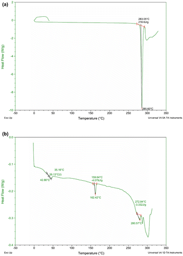
The physical state of 5Fu crystals before and after encapsulation inside PLGA MPs was evaluated using DSC. As the crushed 5Fu powder was suspended in the PLGA-DCM solution during the fabrication of MPs via s/o/w emulsification, it was loaded in its crystalline form by physical entrapment and was found to remain in the same form after encapsulation as shown by DSC thermograms in . The DSC thermograms indicate that the melting point of 5Fu remained more or less the same before ((a)) and after encapsulation inside MPs (b), thus indicating the presence of its crystalline form. Besides the melting peak of 5Fu, the two other small endothermic peaks at 39.1 and 162.4°C in (b) correspond to the T g of PLGA and T m of PLA crystals, respectively.
Figure 10. DSC thermograms showing similar endothermic T m peaks of 5Fu, before and after encapsulation inside PLGA MPs.
Notes: (a) 5Fu powder and (b) 5Fu-loaded 0.4A PLGA MPs.
4.6. X-ray diffraction
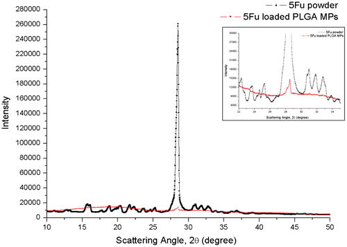
The crystalline nature of 5Fu inside PLGA MPs was determined from XRD spectrum. compares the XRD spectra of 5Fu powder alone and 5Fu-loaded PLGA MPs (the inset is the zoomed out part of the spectrum where 5Fu peak appears). 5Fu powder alone showed a very sharp high intensity peak around 28.5° 2θ corresponding to its crystalline nature. 5Fu-loaded MPs also showed a small peak around 28.5° 2θ, thus indicating the presence of crystalline 5Fu after encapsulation. The smaller magnitude of intensity of 5Fu in case of 5Fu-loaded PLGA MPs is due to the low concentration of 5Fu inside the MPs (part of the 5Fu is also expected to be dissolved in the PLGA matrix). Thus, 5Fu remained in its crystalline form even after encapsulation inside PLGA MPs via s/o/w emulsification.
Figure 11. XRD spectrum of 5Fu powder and 5Fu-loaded 0.4A PLGA MPs indicating crystalline nature of 5Fu before and after encapsulation inside MPs.
Note: The inset shows the overlap of 5Fu intensity peak in both samples over an expanded range of X-axis.
4.7. Nuclear magnetic resonance
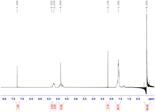
The presence of residual DCM was determined from 1H NMR spectrum for two samples of 5Fu-loaded PLGA MPs (dried for 2 and 4 days). The upper limit of DCM residue in pharmaceutical formulations as approved by US FDA guideline is 600 ppm (parts per million) (Howard, Buttery, Shakesheff, & Roberts, Citation2008; Shelke, Kadam, Tyagi, Rao, & Kompella, Citation2011). NMR spectrum of MPs, dried for 4 days, showed 557 ppm as shown in , while those dried for 2 days showed around 773 ppm of DCM (δ = 5.322 ppm) (Figure S2). Thus, 4 days of drying in vacuum oven at 40°C was sufficient to reduce residual DCM to approved safe limits.
Figure 12. NMR spectrum of 5Fu-loaded PLGA MPs showing safer levels of residual DCM (<600 ppm) after 4 days of drying.
4.8. Syringeability
Syringeability tests were conducted to determine the optimum concentration of HA solution required to develop the unit dose formulation with 5Fu-loaded PLGA MPs. Mass of each 100-μl composite solution sample was weighed to determine if there was any loss through the passage of syringe and needle walls. Different hypodermic needles were used to check which needle would inject the sample without any back pressure and loss in mass of recovered sample. Earlier studies have reported safe use of subconjunctival injections using syringes with needle gage ranging from 25G up to 30G (Chiang et al., Citation2001; How et al., Citation2010; Kozobolis, Konstantinidis, & Labiris, Citation2013; Mead et al., Citation2003; Moritera et al., Citation1991; Perucho-Martínez, Gutiérrez-Díaz, Montero-Rodríguez, Mencía-Gutiérrez, & Lago-Llinás, Citation2006; Peyman et al., Citation1992). Composite solutions, having 1 and 2.3% HA concentration, demonstrated significant back pressure during injection with 25G and 27G needles and there was loss in the recovered sample. Thus, further tests focused on HA with 0.1, 0.25, and 0.5% concentrations.
The composite solutions (100 μl volume) of 5Fu-loaded PLGA MPs with 0.1, 0.25, and 0.5% HA were passed through 1-ml syringe attached to 23G and 25G needles and average mass of injected out MPs was recorded (Table S1). For comparison, the composite solutions were also injected through syringes without any needles to be used as the reference. It was observed that the mass of recovered composite solution after injecting through syringe (with or without needles) remained within a range of 94–104 mg. This indicates that varying HA concentrations for a fixed amount of MPs did not significantly influence the mass of recovered composite solutions.
However, further analysis of amount of 5Fu extracted from these solutions using HPLC showed differences among the various concentrations of HA. In case of 0.1% composite solution, it was observed that the amount of 5Fu present in reference and test samples (injected with 23G and 25G) was significantly (p-value ranging from 0.005 to 0.01) lower than that obtained using 5Fu powder alone (Figure S3). This loss of approximately 50 to 75% 5Fu and inconsistency in amounts could possibly be due to the low viscosity of 0.1% HA solution that results in sedimentation of MPs inside the syringe during injection. The viscosity of HA solution should be optimum to hold the MPs within the polymer chains while allowing their smooth movement and minimizing shear forces across the syringe walls. Visible residues could be observed on the syringe walls indicating 0.1% HA solution was not suitable for the composite formulation.
The composite solutions with 0.25 and 0.5% HA, however, showed better consistency as compared to 0.1% HA in extracted amounts of 5Fu with respect to different gage needles. shows the amount of 5Fu extracted from PLGA MPs injected out from 0.25 to 0.5% HA solutions using 23G and 25G needles. In case of 0.25% HA, the amount of extracted 5Fu was significantly (p-value equal to ~0.003) reduced after using 25G needle in comparison to the reference sample, indicating that decrease of needle gage causes decrease in the mass of recovered composite solution. In case of 0.5% HA, the samples showed better consistency across the varying needle gages. Thus, 25G needle, the smallest gage giving consistent results, was selected as the optimum needle gage for administration of formulation using 0.5% HA solution.
Figure 13. Quantitative analysis of 5Fu extracted from composite solutions having 0.25 and 0.5% HA after injecting them through 1-ml syringe with and without needles (23G and 25G).
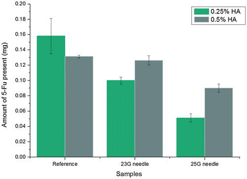
The syringeability and 5Fu quantitative estimation experiments were repeated with 25G needle for composite solution of PLGA MPs in 0.5% HA to further evaluate their reproducibility (Figure S4). Less than 25% loss was observed in the sample recovered from 25G needle. Any difference across experiments (5Fu amounts in recovered sample) could be due to the sedimentation of particles during mixing and differences in drug and size distribution of injected MPs.
4.9. In vitro release
The selected composite formulation of 5Fu-loaded PLGA MPs mixed with 0.5% HA solution was evaluated for in vitro drug release. shows the % cumulative release of 5Fu from PLGA 0.4A MPs alone and after incorporation into 0.5% HA solution. As we can observe, the release followed a sigmoidal pattern and trend was similar in both the samples. There was no difference in the 5Fu release rate between MPs alone and MPs-loaded composite solution indicating that HA chains surrounding the MPs did not provide any barrier to the diffusion of drug.
Figure 14. In vitro release profile of 5Fu from 0.4A PLGA MPs-loaded 0.5% HA solution showing similar rate of release as MPs alone.
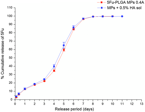
5Fu, being a small molecular weight hydrophilic drug, was not retarded by hydrated or water swollen chains of HA and thus was released with the same rate as that of MPs alone. It diffuses rapidly through HA chains after releasing from entrapped PLGA MPs.
Composite hydrogel systems, obtained by incorporation of 5Fu-loaded PLGA MPs inside HA-MA hydrogels, were also characterized for 5Fu release to investigate if the gel network had any effect on the drug release rate. These gels were prepared as thin disks for release study. shows the % cumulative release of 5Fu from PLGA 0.4A MPs alone and after incorporation into 4% HA-MA hydrogel. As we can observe, the release followed a sigmoidal pattern and trend was similar in both the samples. However, the release was significantly (p-value ranging from 0.001 to 0.01 across days 4 to 10) retarded in composite hydrogel between days 4 and 10, indicating the effect of hydrogel network surrounding the MPs. Also, the composite hydrogel sample released 1–33 μg of 5Fu per day maintaining the therapeutic dosage (1 μg/day) up to 15 days.
Figure 15. In vitro release profile of 5Fu from 0.4A PLGA MPs-loaded 4% HA-MA hydrogel showing retardation in release rate as compared to MPs alone.
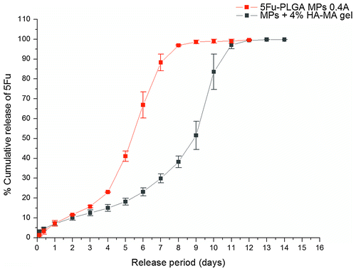
The micron-sized PLGA MPs entrapped inside HA-MA hydrogels were not released outside of gel network that had a mesh size in nanometers. Thus, in composite hydrogels, MPs released the drug while being enclosed within the gel network. The drug diffused through the MPs and then the gel matrix before being released outside into the release medium.
Among various composite hydrogel samples, 4% gels showed maximum retardation. Although there was retardation in release rate for a period of 6 days, the overall release period was extended by 2 days indicating that the HA hydrogel influenced by retarding the 5Fu release from PLGA MPs.
Taken together, our 5-Fu PLGA MPs with HA hydrogel as composite formulation confirm the sustained release of 5Fu for up to 15 days without any burst release. These formulations may provide better localization in the ocular target site compared to only MPs due to highly cross-linked hydrogel network.
5. Conclusion
In this work, 5Fu-loaded PLGA MPs were fabricated and incorporated into HA solution as well as HA hydrogel (HA-MA) to develop composite delivery systems for potential application in ocular anti-fibrotic therapy. Single emulsion s/o/w method was optimized to encapsulate 5Fu inside PLGA MPs and effect of PLGA end group, PLGA concentration, and theoretical drug loading on surface morphology and 5Fu release behavior was investigated. 0.4A PLGA MPs released the drug faster than 0.4E due to faster rate of degradation of the former. Concentration of PLGA affected the size of MPs which in turn affected the encapsulation of 5Fu as well as the practical loading. For a specific size range of MPs, the release rates were not significantly different. Increasing the theoretical loading of 5Fu increased the release rate of drug from PLGA MPs. The batch of MPs with highest practical drug loading was selected for incorporation into HA solution and HA-MA hydrogel and evaluated for syringeability and in vitro release. The release of 5Fu from MPs composite HA-MA hydrogel was significantly retarded up to 15 days without any burst release and these composite HA solution and hydrogel would ensure retention of MPs within the target site for longer periods, thus improving drug bioavailability. The results show that hydrogel composite formulations can be potentially used as drug delivery carriers for therapeutic applications not only for ocular diseases, but also for other diseases as local drug delivery systems.
Acknowledgements
Authors are grateful to Mr. Diels Ding from Fritsch Asia Pacific Pte Ltd, Singapore, for providing the Laser Particle Analyzer facility for size measurement of PLGA microparticles. Authors would also like to acknowledge the generous help and support from Mr. Vishal Mogal and Dr. Zviad Tsakadze, School of Materials Science and Engineering, Nanyang Technological University, Singapore, for the NMR and XRD experiments, respectively.
Additional information
Funding
Notes on contributors
Meghali Bora
The research activities of our group are focused on polymeric delivery systems for biomedical applications. More specifically, it involves modification, design optimization, and characterization of micro/nanoparticulate carriers. Some of the major research areas include localized drug/gene delivery via stents, ocular nanomedicine, shape memory and hemocompatible polymers, cardiac regenerative medicine, injectable implants, and biomedical devices. Our team is closely working with hospitals as well as research centers toward developing therapeutic solutions for various disease targets.
The research work in this paper addresses an important issue of drug delivery. Prolonged sustained drug delivery using biocompatible and biodegradable carriers is highly desirable, as it greatly improves patient compliance. Design and characterization of the formulation reported here make a significant contribution toward the ongoing research activities of ocular drug delivery. Moreover, this work forms a platform for microparticulate ocular delivery systems for further investigation and product development suitable for a wider research audience.
References
- Arias, J. L. (2008). Novel strategies to improve the anticancer action of 5-Fluorouracil by using drug delivery systems. Molecules, 13, 2340–2369.10.3390/molecules13102340
- Bae, J. W., Go, D. H., Park, K. D., & Lee, S. J. (2006). Thermosensitive chitosan as an injectable carrier for local drug delivery. Macromolecular Research, 14, 461–465.10.1007/BF03219111
- Caicco, M. J., Cooke, M. J., Wang, Y., Tuladhar, A., Morshead, C. M., & Shoichet, M. S. (2013). A hydrogel composite system for sustained epi-cortical delivery of cyclosporin a to the brain for treatment of stroke. Journal of Controlled Release, 166, 197–202.10.1016/j.jconrel.2013.01.002
- Chang, E., McClellan, A. J., Farley, W. J., Li, D. Q., Pflugfelder, S. C., & de Paiva, C. S. (2011). Biodegradable PLGA-based drug delivery systems for modulating ocular surface disease under experimental murine dry eye. Journal of Clinical & Experimental Ophthalmology, 2, 191–196.
- Chiang, C. H., Tung, S. H., Lu, D. W., & Yeh, M. K. (2001). In vitro and in vivo evaluation of an ocular delivery system of 5-Fluorouracil microspheres. Journal of Ocular Pharmacology and Therapeutics, 17, 545–553.10.1089/10807680152729239
- Choi, J. S., Seo, K., & Yoo, J. W. (2012). Recent advances in PLGA particulate systems for drug delivery. Journal of Pharmaceutical Investigation, 42, 155–163.10.1007/s40005-012-0024-5
- Cook, C., & Foster, P. (2012). Epidemiology of glaucoma: What’s new? Canadian Journal of Ophthalmology, 47, 223–226.10.1016/j.jcjo.2012.02.003
- Cui, L., Sun, N., Li, X., Huang, J., & Yang, J. (2008). Subconjunctival sustained release 5-fluorouracil for glaucoma filtration surgery. Acta Pharmacologica Sinica, 29, 1021–1028.10.1111/aphs.2008.29.issue-9
- Faisant, N., Siepmann, J., & Benoit, J. P. (2002). PLGA-based microparticles: Elucidation of mechanisms and a new, simple mathematical model quantifying drug release. European Journal of Pharmaceutical Sciences, 15, 355–366.10.1016/S0928-0987(02)00023-4
- Fredenberg, S., Wahlgren, M., Reslow, M., & Axelsson, A. (2011). The mechanisms of drug release in poly(lactic-co-glycolic acid)-based drug delivery systems–A review. International Journal of Pharmaceutics, 415, 34–52.10.1016/j.ijpharm.2011.05.049
- Galeska, I., Kim, T. K., Patil, S. D., Bhardwaj, U., Chatttopadhyay, D., Papadimitrakopoulos, F., & Burgess, D. J. (2005). Controlled release of dexamethasone from PLGA microspheres embedded within polyacid-containing PVA hydrogels. The AAPS Journal, 7, E231–E240.10.1208/aapsj070122
- Gooch, N., Molokhia, S. A., Condie, R., Burr, R. M., Archer, B., Ambati, B. K., & Wirostko, B. (2012). Ocular drug delivery for glaucoma management. Pharmaceutics, 4, 197–211.10.3390/pharmaceutics4010197
- Green, E., Wilkins, M., Bunce, C., & Wormald, R. (2014). 5-Fluorouracil for glaucoma surgery. The Cochrane Database of Systematic Reviews,(2), 1–62. doi:10.1002/14651858.CD001132.pub2
- Hoare, T. R., & Kohane, D. S. (2008). Hydrogels in drug delivery: Progress and challenges. Polymer, 49, 1993–2007.10.1016/j.polymer.2008.01.027
- Hou, Q., Chau, D. Y., Pratoomsoot, C., Tighe, P. J., Dua, H. S., Shakesheff, K. M., & Rose, F. R. (2008). In situ gelling hydrogels incorporating microparticles as drug delivery carriers for regenerative medicine. Journal of Pharmaceutical Sciences, 97, 3972–3980.10.1002/jps.21310
- How, A., Chua, J. L. L., Charlton, A., Su, R., Lim, M., Kumar, R. S., … Wong, T. T. (2010). Combined treatment with bevacizumab and 5-fluorouracil attenuates the postoperative scarring response after experimental glaucoma filtration surgery. Investigative Ophthalmology & Visual Science, 51, 928–932.
- Howard, D., Buttery, L. D., Shakesheff, K. M., & Roberts, S. J. (2008). Tissue engineering: Strategies, stem cells and scaffolds. Journal of Anatomy, 213, 66–72.10.1111/joa.2008.213.issue-1
- Huhtala, A. R. S., Rönkkö, S., Teräsvirta, M., Puustjärvi, T., Sihvola, R., Vehanen, K., … Uusitalo, H. (2009). The effects of 5-fluorouracil on ocular tissues in vitro and in vivo after controlled release from a multifunctional implant. Investigative Opthalmology & Visual Science, 50, 2216–2223.10.1167/iovs.08-3016
- Jacob, J. T., LaCour, O. J., & Burgoyne, C. F. (2001). Slow release of the antimetabolite 5-fluorouracil (5-FU) from modified Baerveldt glaucoma drains to prolong drain function. Biomaterials, 22, 3329–3335.10.1016/S0142-9612(01)00170-3
- Jin, Y. J., Ubonvan, T., & Kim, D. D. (2010). Hyaluronic acid in drug delivery systems. Journal of Pharmaceutical Investigation, 40, 33–43.10.4333/KPS.2010.40.S.033
- Joung, Y. K., Choi, J. H., Park, K. M., & Park, K. D. (2007). PLGA microparticle-embedded thermosensitive hydrogels for sustained release of hydrophobic drugs. Biomedical Materials, 2, 269–273.10.1088/1748-6041/2/4/010
- Ju, R., Wen, Y., Gou, R., Wang, Y., & Xu, Q. (2014). The experimental therapy on brain ischemia by improvement of local angiogenesis with tissue engineering in the mouse. Cell Transplantation, 23, S83–S95.10.3727/096368914X684998
- Kimura, H., & Ogura, Y. (2001). Biodegradable polymers for ocular drug delivery. Ophthalmologica, 215, 143–155.10.1159/000050849
- Kingman, S. (2004). Glaucoma is second leading cause of blindness globally. Bulletin of the World Health Organization, 82, 887–888.
- Kogan, G., Soltes, L., Stern, R., & Gemeiner, P. (2007). Hyaluronic acid: A natural biopolymer with a broad range of biomedical and industrial applications. Biotechnology Letters, 29, 17–25.
- Kondo, M., & Aroie, M. (1989). Iontophoresis of 5-fluorourocil into the conjunctiva ond sclero. Investigative Ophthalmology & Visual Science, 30, 583–585.
- Kozobolis, V., Konstantinidis, A., & Labiris, G. (2013). Combined cataract-glaucoma surgery. In: S. Rumelt (Ed.) Glaucoma–basic and clinical aspects.(pp. 473–510). Israel: InTech.
- Lagarce, F., Faisant, N., Desfontis, J. C., Marescaux, L., Gautier, F., Richard, j., … Benoit, J. P. (2005). Baclofen-loaded microspheres in gel suspensions for intrathecal drug delivery: In vitro and in vivo evaluation. European Journal of Pharmaceutics and Biopharmaceutics, 61, 171–180.10.1016/j.ejpb.2005.04.004
- Lee, D. A., Hersh, P., Kersten, D., & Melamed, S. (1987). Complications of subconjunctival 5-fluorouracil following glaucoma filtering surgery. Ophthalmic Surgery, 18, 187–190.
- Lin, Y., Sun, J., Jiang, G., Zan, J., & Ding, F. (2007). In vitro evaluation of lysozyme-loaded microspheres in methylcellulose hydrogel. Chinese Journal of Chemical Engineering, 15, 566–572.10.1016/S1004-9541(07)60125-6
- Longley, D. B., Harkin, D. P., & Johnston, P. G. (2003). 5-Fluorouracil: Mechanisms of action and clinical strategies. Nature Reviews Cancer, 3, 330–338.10.1038/nrc1074
- Lu, D. W., Chang, C. J., Chiang, C. H., Yeh, M. K., & Chou, P. I. (2000). Wound modulation after trabeculectomy by different formulations of antimetabolites in rabbits. Journal of Ocular Pharmacology and Therapeutics, 16, 529–538.10.1089/jop.2000.16.529
- Mahoney, B. P., Raghunand, N., Baggett, B., & Gillies, R. J. (2003). Tumor acidity, ion trapping and chemotherapeutics I. Acid pH affects the distribution of chemotherapeutic agents in vitro. Biochemical Pharmacology, 66, 1207–1218.10.1016/S0006-2952(03)00467-2
- Makadia, H. K., & Siegel, S. J. (2011). Poly lactic-co-glycolic acid (PLGA) as biodegradable controlled drug delivery carrier. Polymers, 3, 1377–1397.10.3390/polym3031377
- Mead, A. L., Wong, T. T. L., Cordeiro, M. F., Anderson, I. K., & Khaw, P. T. (2003). Evaluation of anti-TGF-2 antibody as a new postoperative anti-scarring agent in glaucoma surgery. Investigative Ophthalmology & Visual Science, 44, 3394–3401.
- Moritera, T., Ogura, Y., Honda, Y., Wada, R., Hyon, S. H., & Ikada, Y. (1991). Microspheres of biodegradable polymers as a drug-delivery system in the vitreous. Investigative Ophthalmology & Visual Science, 32, 1785–1790.
- Mufamadi, M. S., Pillay, V., Choonara, Y. E., Du Toit, L. C., Modi, G., Naidoo, D., & Ndesendo, V. M. (2011). A review on composite liposomal technologies for specialized drug delivery. Journal of Drug Delivery, 2011, 1–19.10.1155/2011/939851
- Mundargi, R. C., Babu, V. R., Rangaswamy, V., Patel, P., & Aminabhavi, T. M. (2008). Nano/micro technologies for delivering macromolecular therapeutics using poly(D,L-lactide-co-glycolide) and its derivatives. Journal of Controlled Release, 125, 193–209.10.1016/j.jconrel.2007.09.013
- Naguib, Y. W., Kumar, A., & Cui, Z. (2014). The effect of microneedles on the skin permeability and antitumor activity of topical 5-fluorouracil. Acta Pharmaceutica Sinica B, 4, 94–99.10.1016/j.apsb.2013.12.013
- Nasr, M., Ghorab, M. K., & Abdelazem, A. (2015). In vitro and in vivo evaluation of cubosomes containing 5-fluorouracil for liver targeting. Acta Pharmaceutica Sinica B, 5, 79–88.10.1016/j.apsb.2014.12.001
- Nath, S. D., Linh, N. T., Sadiasa, A., & Lee, B. T. (2014). Encapsulation of simvastatin in PLGA microspheres loaded into hydrogel loaded BCP porous spongy scaffold as a controlled drug delivery system for bone tissue regeneration. Journal of Biomaterials Applications, 28, 1151–1163.10.1177/0885328213499272
- Nettles, D. L., Vail, T. P., Morgan, M. T., Grinstaff, M. W., & Setton, L. A. (2004). Photocrosslinkable hyaluronan as a scaffold for articular cartilage repair. Annals of Biomedical Engineering, 32, 391–397.10.1023/B:ABME.0000017552.65260.94
- Patterson, J., Stayton, P. S., & Li, X. (2009). In situ characterization of the degradation of PLGA microspheres in hyaluronic acid hydrogels by optical coherence tomography. IEEE Transactions on Medical Imaging, 28, 74–81.10.1109/TMI.2008.927356
- Perucho-Martínez, S., Gutiérrez-Díaz, E., Montero-Rodríguez, M., Mencía-Gutiérrez, E., & Lago-Llinás, M. D. (2006). Needle revision of late failing filtering blebs after glaucoma surgery. Archives of the Spanish Society of Ophthalmology, 81, 517–522.
- Peyman, G. A., Conway, M., Khoobehi, B., & Soike, K. (1992). Clearance of microsphere-entrapped 5-fluorouracil and cytosine arabinoside from the vitreous of primates. International Ophthalmology, 16, 109–113.10.1007/BF00918942
- Quigley, H. A. (2011). Glaucoma. The Lancet, 377, 1367–1377.10.1016/S0140-6736(10)61423-7
- Quigley, H. A., & Borman, A. T. (2006). The number of people with glaucoma worldwide in 2010 and 2020. British Journal of Ophthalmology, 90, 262–267.10.1136/bjo.2005.081224
- Ranganath, S. H., Kee, I., Krantz, W. B., Chow, P. K., & Wang, C. H. (2009). Hydrogel matrix entrapping PLGA-paclitaxel microspheres: Drug delivery with near zero-order release and implantability advantages for malignant brain tumour chemotherapy. Pharmaceutical Research, 26, 2101–2114.10.1007/s11095-009-9922-2
- Rocíoh, V. (2011). Microparticles as drug delivery systems for the back of the eye. In B. E. H. F. Kompellau (Ed.), Drug product development for the back of the eye (pp. 231–259). New York, NY: American Association of Pharmaceutical Scientists.
- Samadi, N., Abbadessa, A., Di Stefano, A., van Nostrum, C. F., Vermonden, T., Rahimian, S., … Hennink, W. E. (2013). The effect of lauryl capping group on protein release and degradation of poly(D,L-lactic-co-glycolic acid) particles. Journal of Controlled Release, 172, 436–443.10.1016/j.jconrel.2013.05.034
- Shelke, N. B., Kadam, R., Tyagi, P., Rao, V. R., & Kompella, U. B. (2011). Intravitreal poly(L-lactide) microparticles sustain retinal and choroidal delivery of TG-0054, a hydrophilic drug intended for neovascular diseases. Drug Delivery and Translational Research, 1, 76–90.10.1007/s13346-010-0009-8
- Siegelr, A., & Rathbonem, J. (2012). Overview of controlled release mechanisms. In J. Siepmann, A. Siegelr, & J. Rathbonem (Eds.), Fundamentals and applications of controlled release drug delivery, advances in delivery science and technology. (pp. 19–43). New York, NY: Springer.
- Smeds, K. A., Pfister-Serres, A., Miki, D., Dastgheib, K., Inoue, M., Hatchell, D. L., & Grinstaff, M. W. (2000). Photocrosslinkable polysaccharides for in situ hydrogel formation. Journal of Biomedical Materials Research, 54, 115–121.
- Tous, E., Weber, H. M., Lee, M. H., Koomalsingh, K. J., Shuto, T., Kondo, N., … Burdick, J. A. (2012). Tunable hydrogel-microsphere composites that modulate local inflammation and collagen bulking. Acta Biomaterialia, 8, 3218–3227.10.1016/j.actbio.2012.05.027
- Wang, G., Tucker, I. G., Roberts, M. S., & Hirst, L. W. (1996). In vitro and in vivo evaluation in rabbits of a controlled release 5-fluorouracil subconjunctival implant based on poly(D,L-lactide-co-glycolide). Pharmaceutical Research, 13, 1059–1064.10.1023/A:1016062825360
- Wang, Y., Wei, Y. T., Zu, Z. H., Ju, R. K., Guo, M. Y., Wang, X. M., … Cui, F. Z. (2011). Combination of hyaluronic acid hydrogel scaffold and PLGA microspheres for supporting survival of neural stem cells. Pharmaceutical Research, 28, 1406–1414.10.1007/s11095-011-0452-3
- Widjaja, L. K., Bora, M., Chan, P. N., Lipik, V., Wong, T. T., & Venkatraman, S. S. (2014). Hyaluronic acid-based nanocomposite hydrogels for ocular drug delivery applications. Journal of Biomedical Materials Research Part A, 102, 3056–3065.10.1002/jbm.a.v102.9
- Wormald, R., Wilkins, M., & Bunce, C. (2001). Post-operative 5-fluorouracil for glaucoma surgery. The Cochrane Database of Systematic Reviews,(3), 1–33. doi:10.1002/14651858.CD001132.pub2
- Yeh, M. K., Tung, S. M., Lu, D. W., Chen, J. L., & Chiang, C. H. (2001). Formulation factors for preparing ocular biodegradable delivery system of 5-fuorouracil microparticles. Journal of Microencapsulation, 18, 507–519.
- You, H., Lee, E. U., Kim, Y. K., Kim, B. C., Park, J. Y., Lim, H. C., … Choi, S. H. (2014). Biocompatibility and resorption pattern of newly developed hyaluronic acid hydrogel reinforced three-layer poly (lactide-co-glycolide) membrane: Histologic observation in rabbit calvarial defect model. Biomaterials Research, 18, 12–18.10.1186/2055-7124-18-12
- Zhao, J., Guo, B., & Ma, P. X. (2014). Injectable alginate microsphere/PLGA–PEG–PLGA composite hydrogels for sustained drug release. RSC Advances, 4, 17736–17742.10.1039/c4ra00788c
- Zimmer, A., & Kreuter, J. (1995). Microspheres and nanoparticles used in ocular delivery systems. Advanced Drug Delivery Reviews, 16, 61–73.10.1016/0169-409X(95)00017-2