Abstract
Congenital heart disease (CHD) is the most common congenital birth defect, and hypoplastic left heart syndrome (HLHS) is a relatively common form of CHD, with an estimated incidence of 0.1–0.25 per 1,000 live births. However, twin pregnancies in which both fetuses are affected by HLHS are very rare. Herein, we present the first reported case of dichorionic dizygotic twins concordant for this condition.
Public Interest Statement
Congenital heart disease is the most common congenital birth defect. However twin pregnancies in which both fetuses are affected by hypoplastic left heart syndrome are very rare. Herein, we present the first reported case of dichorionic dizygotic twins concordant for this condition.
Competing Interests
The authors declare no competing interest.
1. Introduction
Congenital heart disease (CHD) is the most common congenital birth defect and the leading cause of infant morbidity and disability, with an incidence rate of 6–8 per 1,000 live births (Hoffman & Kaplan, Citation2002). Hypoplastic left heart syndrome (HLHS) is a relatively common form of CHD, with an estimated incidence rate of 0.1–0.25 per 1,000 live births (Abu-Harb, Hey, & Wren, Citation1994). HLHS refers to congenital hypoplasia of the left ventricle that may be associated with mitral atresia, aortic atresia, aortic stenosis, and coarctation of the aorta (Simpson, Citation2000). In patients with this condition, the heart is not able to maintain systemic circulation. While the severity of this defect can vary, it is generally fatal if left untreated. HLHS is diagnosed sonographically with a four-chamber view, usually at 18–22 weeks of gestation. There have been previous reports of HLHS in monochorionic twin pregnancies affecting one fetus (Dumontier, Fermont, Le Bidois, et al., Citation2002; Negishi, Okuyama, Sagawa, Makinoda, & Fujimoto, Citation1995) or both fetuses (Andrews et al., Citation2004), but to our knowledge, this is the first case of dichorionic dizygotic twins concordant for this condition.
2. Case report
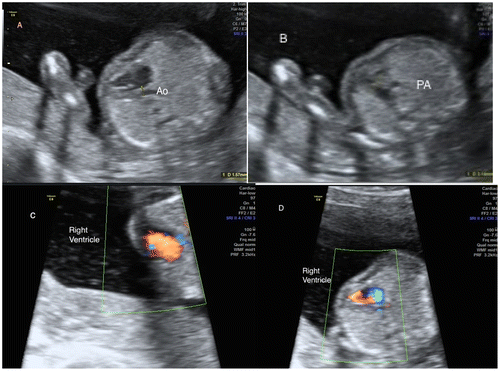
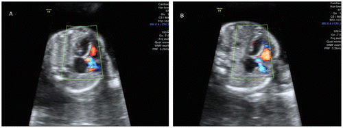
In this report, we describe the case of a dichorionic, diamniotic, dizygotic twin pregnancy in which both fetuses were diagnosed with HLHS by antenatal sonography at 17 weeks’ gestation. This was the first pregnancy for the mother, who was 31 years of age. She had no chronic illnesses such as diabetes, hypertension, asthma, or obesity, and no alcohol or smoking habits. She had anemia, with an Hb level of 9.6 g/dL. On her first ultrasound examination at 13 weeks’ gestation, nuchal translucency was 2.0 mm in both fetuses, and no major abnormalities were seen. In a fetal sonography taken at 17 weeks’ gestation, the cardiac left ventricle of the female fetus was hypoplastic; the diameter of the aorta was 1.57 mm and that of the pulmonary artery was 3.14 mm. Color Doppler sonography revealed that there was no blood flow to the left ventricle (). Also at 17 weeks’ gestation, the male fetus had a normal four-chamber view, but the color Doppler assessment showed no blood flow to the left ventricle and severe mitral valve stenosis (). Both pulmonary and systemic circulation were dependent on the right ventricle, with flow to the aorta being provided through a patent arterial duct. The family, faced with a very difficult decision, ultimately elected to terminate the pregnancy, which was performed by inducing labor. Samples were collected from both fetuses for genetic examination, and the karyotype analyses for both fetuses were normal (46, XX and 46, XY).
3. Discussion
Over the past several decades, ultrasonography and fetal echocardiography have enabled the identification of HLHS in utero, and through improvements in ultrasound resolution, this diagnosis can be made as early as 12–14 weeks’ gestation. This advancement allows parents time to learn about this condition and to consider the option of terminating the pregnancy. If termination is chosen, early diagnosis helps reduce psychological trauma and maternal complications. If the parents choose to continue the pregnancy, the prenatal diagnosis allows the health care professional to plan perinatal management and to perform further evaluations for abnormal karyotypes. Our patient chose to terminate the pregnancy, after which karyotype evaluations were performed on both fetuses.
There have been several suggested causes for the manifestation of HLHS. An early study suggested that it was an immunoreactive response by the heart tissues to specific growth factors: the heart tissues under- or overreact to the presence of growth factors and are unable to form or grow properly (Burton et al., Citation1991). Further research has indicated that there is a genetic component to this defect; families with other known cardiac defects are more likely to have offspring with HLHS (Cronk, Pelech, Malloy, & McCarver, Citation2004). Although the risk of structural heart disease is higher in twin pregnancies than in singleton pregnancies, the concordance rate is relatively low in monochorionic pregnancies (Dumontier et al., Citation2002), implicating non-genetic factors in the etiology. However, a previous case report described a case of monochorionic twins concordant for HLHS (Andrews et al., Citation2004), suggesting that at least in some cases, there may be a genetic contribution to the etiology. To our knowledge, the present report is the first case of dichorionic dizygotic twins concordant for HLHS, which would indicate a genetic contribution to the etiology. However, the karyotypes of these fetuses were normal. While both fetuses had HLHS, they had different morphologic anomalies. The female fetus only had a hypoplastic left ventricle, and outflow of the ventricle was normal. In the male fetus, the HLHS was due to mitral stenosis. Recent studies have shown that the rate of CHD increases in siblings (Boughman et al., Citation1987; Hoess, Goldmuntz, & Pyeritz, Citation2002; Mu, McAdams, & Bush, Citation2005; Oyen et al., Citation2009; Wang, Wang, & Zhao, Citation2013). A population-based epidemiologic study calculated a 13.5% sibling recurrence risk for right- or left-sided flow lesions in probands with HLHS (Boughman et al., Citation1987; Mu et al., Citation2005).
HLHS is the most severe form of CHD, and until recently it was considered to be fatal. HLHS carries a poor prognosis, with a reported 40–50% survival rate after prenatal diagnosis compared with neonates born with HLHS that was not diagnosed before birth (Allan, Apfel, & Printz, Citation1998; Brackley et al., Citation2000). However, surgical intervention is now possible using neonatal heart transplantation or three-stage palliative surgery (Norwood procedure). Nevertheless, HLHS remains a leading cause of infant death due to cardiovascular malformation and is associated with significant long-term morbidity.
A twin pregnancy is considered to be one of the risk factors leading to CHD, and the concordance rate of monozygotic twins is higher than that of dizygotic twins (Taylor & Fisk, Citation2000). In this report, we described the case of dichorionic twins concordant for this manifestation. Both genetic and environmental factors may be important in the etiology of HLHS.
Additional information
Funding
Notes on contributors
Halis Özdemir
Başkent University is the first private university to teach health sciences in Turkey and I am a member of perinatolgy unit of Baskent University Medical School Adana Teaching and Medical Research Center, Department of Obstetrics & Gynecology. We work as a team and working consultant physician in the field of perinatology for Çukurova region in Turkey.
References
- Abu-Harb, M., Hey, E., & Wren, C. (1994). Death in infancy from unrecognised congenital heart disease. Archives of Disease in Childhood, 71, 3–7.10.1136/adc.71.1.3
- Allan, L. D., Apfel, H. D., & Printz, B. F. (1998). Outcome after prenatal diagnosis of the hypoplastic left heart syndrome. Heart, 79, 371–373.10.1136/hrt.79.4.371
- Andrews, R. E., Yates, R. W. M., Sullivan, I. D., Cook, A. C., Anderson, R. H., & Lees, C. C. (2004). Early fetal diagnosis of monochorionic twins concordant for hypoplastic left heart syndrome. Ultrasound in Obstetrics and Gynecology, 24, 101–102.10.1002/(ISSN)1469-0705
- Boughman, J. A., Berg, K. A., Astemborski, J. A., Clark, E. B., McCarter, R. J., Rubin, J. D., … Reynolds, J. F. (1987). Familial risks of congenital heart defect assessed in a population-based epidemiologic study. American Journal of Medical Genetics, 26, 839–849.10.1002/(ISSN)1096-8628
- Brackley, K. J., Kilby, M. D., Wright, J. G., Brawn, W. J., Sethia, B., Stumper, O., … Whittle, M. J. (2000). Outcome after prenatal diagnosis of hypoplastic left-heart syndrome: a case series. The Lancet, 356, 1143–1147.10.1016/S0140-6736(00)02756-2
- Burton, P. B. J., Hauck, A., Nehlsen-Cannarella, S. L., Gusewitch, G. A., Sorensen, C. M., Gundry, S. R., & Bailey, L. L. (1991). Hypoplastic left heart syndrome: some clues to its aetiology. The Lancet, 338, 1148.10.1016/0140-6736(91)92006-N
- Cronk, C. E., Pelech, A. N., Malloy, M. E., & McCarver, D. G. (2004). Excess birth prevalence of hypoplastic left heart syndrome in Eastern Wisconsin for birth cohorts 1997-1999. Birth Defects Research Part A: Clinical and Molecular Teratology, 70, 114–120.10.1002/(ISSN)1542-0760
- Dumontier, J., Fermont, L., Le Bidois, J., et al. (2002). Abstracts and posters of the XXXVII Annual Meeting of The Association for European Paediatric Cardiology, Porto, 15–18 May, 2002. Cardiology in the Young, 12(S1), 1–87. doi:10.1017/S104795110001218X
- Hoess, K., Goldmuntz, E., & Pyeritz, R. E. (2002). Genetic counseling for congenital heart disease: New approaches for a new decade. Current Cardiology Reports, 4, 68–75.10.1007/s11886-002-0129-y
- Hoffman, J. L., & Kaplan, S. (2002). The incidence of congenital heart disease. Journal of the American College of Cardiology, 39, 1890–1900.10.1016/S0735-1097(02)01886-7
- Mu, T. S., McAdams, R. M., & Bush, D. M. (2005). A case of hypoplastic left heart syndrome and bicuspid aortic valve in monochorionic twins. Pediatric Cardiology, 26, 884–885.10.1007/s00246-005-1016-2
- Negishi, H., Okuyama, K., Sagawa, T., Makinoda, S., & Fujimoto, S. (1995). Two cases of monozygotic twins, in each of which one fetus was prenatally diagnosed as having a heart anomaly. Journal of Obstetrics and Gynaecology, 21, 293–298.10.1111/(ISSN)1447-0756c
- Oyen, N., Poulsen, G., Boyd, H. A., Wohlfahrt, J., Jensen, P. K., & Melbye, M. (2009). Recurrence of congenital heart defects in families. Circulation, 120, 295–301.10.1161/CIRCULATIONAHA.109.857987
- Simpson, J. M. (2000). Hypoplastic left heart syndrome. Ultrasound in Obstetrics and Gynecology, 15, 271–278.10.1046/j.1469-0705.2000.00086.x
- Taylor, M. J., & Fisk, N. M. (2000). Prenatal diagnosis in multiple pregnancy. Best Practice & Research Clinical Obstetrics & Gynaecology, 14, 663–675.10.1053/beog.2000.0103
- Wang, X., Wang, J., & Zhao, P. (2013). Familial congenital heart disease: Data collection and preliminary analysis. Cardiology in the Young, 23, 394–399.10.1017/S1047951112001035


