Abstract
Background: Increased anterior pelvic tilt is one important contributor to poor posture in children and adolescents and caused by muscular imbalance. This study aims at identifying the extent to which a sensorimotor training reduces anterior pelvic tilt more effectively than pure strength and stretch training alone. Methods: 54 male adolescents (age 13–17) with an increased pelvic tilt angle >14° were matched to three groups (strength training STR, strength + sensorimotor training STR + SENS, control CON). Maximum isometric torques for knee flexion (HA), trunk flexion (RA), and trunk extension, and pelvic tilt were measured before and after a 12 week physical therapy schedule. Two-way mixed ANOVA were calculated. Results: For STR and STR + SENS the relative torque of HA and RA increased significantly (p < 0.05) between pre- and post-test. Significant improvement of the pelvic tilt angle was identified in both training groups, with STR + SENS exhibiting a significantly larger degree of improvement. Conclusions: Sensorimotor exercises improve the effectiveness of physical fitness training to reduce anterior pelvic tilt and should therefore supplement existing therapeutic programs.
Public Interest Statement
More than 60% of all adolescents exhibit weakness in posture, such as hyperlordosis (hollow back). Very often, this leads to back problems. Weak or inelastic muscles result in the pelvic being tilted forward and thus creating a hollow back. In our study, we examined whether targeted gym training of specific muscle groups can help improve posture in adolescents. To do so, we analyzed strength and stretch training as well as special exercises that improve body awareness. We were able to prove that combined training consisting of strength, stretching, and body awareness exercises effectively ameliorates body posture. Specific gym training helps adolescents to prevent or correct poor posture.
Competing Interests
The authors declare no competing interest.
1. Introduction
Between 22 and 65% of children and adolescents demonstrate poor posture (Bogdanović & Marković, Citation2010; Gh, Alilou, Ghafurinia, & Fereydounnia, Citation2012; Kopecký, Citation2004). Even if current studies on the correlation between posture deficits and back pain do not all agree (Laird, Gilbert, Kent, & Keating, Citation2014; Nourbakhsh & Arab, Citation2002; Smith, O'Sullivan, & Straker, Citation2008) and the long-term consequences of poor posture in adolescence have not been clearly established, individual posture characteristics are categorized as health-relevant (Chaléat-Valayer et al., Citation2011; Dolphens, Cagnie, & Coorevits, Citation2012). An increased anterior pelvic tilt is one key cause of hyperlordosis of the lumbar spine and thus a posture-constituting characteristic of therapeutic importance (Le Huec, Aunoble, Philippe, & Nicolas, Citation2011; Roussouly & Pinheiro-Franco, Citation2011). Hyperlordosis of the lumbar spine (“hollow back”) is a typical characteristic of poor posture with possible negative health implications (Jentzsch et al., Citation2013; Smith et al., Citation2008). Therefore, preventive therapeutic treatment of an increased pelvic tilt or lumbar hyperlordosis is important for adolescents.
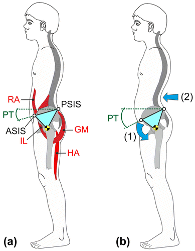
One conceivable reason for an increased anterior pelvic tilt is assumed to be an imbalance of the muscles that stabilize the pelvis (Buchtelová, Tichy, & Vaniková, Citation2013). The orientation of the pelvis in the sagittal plane is explained by a “see-saw model” with the pivot point located in the hip joints ((a)). Depending on the activities of the relevant muscles and the structure of the ligamentous apparatus and the fasciae, the pelvic tilt changes (Alvim, Peixoto, Vicente, Chagas, & Fonseca, Citation2010; Kim et al., Citation2006; López-Miñarro, Muyor, Belmonte, & Alacid, Citation2012). Due to muscular imbalance, the pelvis tilts forward and lumbar lordosis increases. When the thoracic spine compensates for this malposition, an increased thoracal kyphosis and typical posture weakness develop ((b)). Strength and stretch exercises have been proven effective as therapeutic interventions for the muscle groups involved (Page, Citation2006). Earlier studies proved that targeted strength programs for the muscle groups that reduce pelvic tilt resulted in an improvement of posture in adolescents with increased anterior pelvic tilt (Klee, Citation1994). Therefore, the correction of pelvic tilt is a crucial therapeutic goal to improve posture weakness in adolescents.
Figure 1. (a) Pivot model of anterior pelvic tilt, RA–rectus abdominis, IL–iliopsoas, GM–gluteus maximus, HA–hamstrings, PA–pelvic tilt angle, ASIS–anterior superior iliac spine, PSIS–posterior superior iliac spine. (b) Muscular imbalance leads to an increased anterior pelvic tilt (1) and an increased lumbar lordosis (2).
In the past few years, the importance of sensorimotor exercises in fitness training and physical therapy to treat neuromuscular deficits has been emphasized. Sensorimotor training is normally understood as a balance training during which the body position has to be stabilized on moveable auxiliary equipment, thus enhancing the reflective activation of the deep postural muscles (McCaskey, Schuster-Amft, Wirth, Suica, & de Bruin, Citation2014). Therapeutic experience in pediatrics shows that it is important to teach adolescents with poor posture a clear idea of movement, i.e. how to correct their posture especially in regard to the pelvis (Page, Citation2006). However, uniform evidence on this aspect is still lacking.
This study aims to identify the extent to which a combination of strength and stretch training and sensorimotor training might result in a more effective reduction of anterior pelvic tilt in a standing position than pure strength and stretch training.
2. Methods
2.1. Participants
The subjects were included during an interdisciplinary research project (Kid-Check) in 2013 and 2014 by means of newspaper ads. The study included male adolescents with an increased pelvic tilt (anteversion angle >14°). A pelvic tilt between 5° and 13° is thought to be normal for male adolescents (Nguyen & Shultz, Citation2007). Thus, from a preventive point of view, all the subjects included in the study should benefit from a preventive therapy concept. Within the framework of an orthopedic examination of the postural and musculoskeletal system, 54 of 69 interested persons were identified who did not have any spinal deformities or other orthopedic problems, whose body mass index was between 16 and 24, and who did not actively perform any sports at the time. A total of 54 male adolescents between 13 and 17 years having reached a physical development level of at least Tanner IV participated in the study (). A well-matched study design was used in order to minimize possible confounding effects like age and body weight. Initially, all participants were ranked with the pelvic tilt angle being the primary, age the secondary, and body weight the third criterion. Then the first three on the ranking list were randomly assigned to the three groups (resistance training, resistance + senso, control). This process was repeated respectively for three participants until all 54 adolescents were matched to the three groups.
Table 1. Anthropometric data of the participants
Each subgroup consisted of 18 subjects. One subgroup carried out pure stretch and strength training (STR), the other subgroup added body perception training (STR + SENSO), the third group served as a non-exercising control group (CON).
The study’s concept based on the Helsinki declaration. The local ethics commission had approved the study (Ethics Committee of the Saarland University, Prof. Dr C. König, Ref. 15–6). The test persons and their parents had been informed on the order of the study and the content of the training and therapeutic intervention and had given their written consent.
2.2. Measures
The anterior superior iliac spines (ASIS) and posterior superior iliac spines (PSIS) were palpated by an experienced tester and marked with self-adhesive marker balls (diameter: 12 mm). During the examination, the test persons were photographed wearing bathing suits and standing in a casual pose. The test persons were instructed to assume a relaxed pose, look straight ahead, and keep their arms to the sides of their body ().
The anteversion angle of the pelvis (connecting line between the PSIS and ASIS markers against the horizontal line) was determined using specific software (Corpus Concepts, AFG, Idar-Oberstein, Germany). The reliability of this measurement method to determine the pelvic tilt had been proven in earlier studies (Gajdosik, Simpson, Smith, & DonTigny, Citation1985).
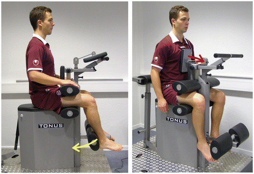
Mainly the straight abdominal muscles (M. rectus abdominis) as trunk flexor, the gluteal muscles (M. gluteus maximus) as a hip extensor and M. biceps femoris as a hip extensor are responsible for a retroversion (straightening up) of the pelvis. Their activity leads to a decrease of pelvic tilt (Buchtelová et al., Citation2013). The maximum isometric torque for knee flexion, trunk flexion, and trunk extension were measured in a sitting position using an isometric measurement device (Easytorque, Tonus Inc., Zemmer, Germany). The knee angle was set to 80° for measuring the knee flexion; the hip angle was set to 90°. Hip and thigh were fixed by means of pads (, left). For measuring the trunk flexion, the trunk was fixed by means of a pad at the sternum’s center height (, right), and for trunk extension, a pad stabilized the trunk at the thoracolumbar region. The test persons were instructed to build up strength for five seconds and hold it for five more. For familiarization purposes, a test trial was performed for each measurement. After that, three isometric tests were executed. Whenever the third value turned out to be the maximum value, further measurements were added until the last measurement value was smaller than the penultimate value. The maximum value reached was then divided by the current body weight and processed further.
2.3. Physical fitness program
The adolescents exercised after their initial examination for 12 weeks twice a week under supervision of an experienced trainer. After a six-min warm-up phase on a treadmill, they performed the strength-endurance exercises in the form of a set-of-three training at different training devices (15 repetitions, 1 min rest). The weights were chosen so that the participants were able to carry out the three sets without errors, but feeling definitely exhausted afterwards (Borg scale 7 of 10). The stretch exercises (active antagonist contract stretching (Ryan, Rossi, & Lopez, Citation2010)) were executed three times each for 40 s for each side of the body. The training included strength exercises for M. rectus abdominis (RA), M. gluteus maximus (GM), and M. biceps femoris (HA) (), as well as stretching exercises for M. rectus femoris and M. iliopsoas, since connections between the extensibility of these muscles (increased range of motion) and the pelvic tilt are being discussed (Bridger, Orkin, & Henneberg, Citation1992; López-Miñarro et al., Citation2012).
Figure 3. Strength training on the machine.
Note: Left: Seated crunch machine (rectus abdominis), middle: knee curl machine (biceps femoris), right: hip extension machine (gluteus maximus).
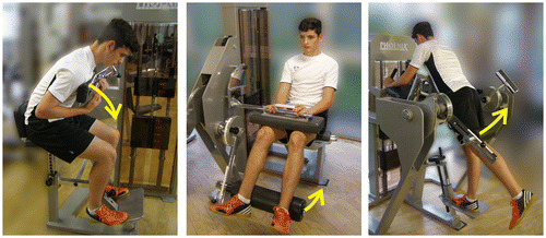
The subgroup that performed body perception training executed four additional exercises () under supervision of an experienced physical therapist. The focus of the additional program was on perception of the pelvis position with open and closed eyes, conscious pelvic retroversion, as well as adjustment of lumbar lordosis in a lying position. The additional program took approximately 20 min and was carried out before the actual strength training because Bruhn, Kullmann, and Gollhofer (Citation2006) were able to observe positive preconditioning effects of sensorimotor training.
Figure 4. Exercises to improve body perception in the pelvic region. (a) Pelvis lift in a lying position: lifting the pelvis a few centimeters, (b) elimination of lumbar lordosis: drawing in the navel, (c) retroversion of the pelvis: turn the pelvis backwards like a wheel, (d) controlling pelvis and posture in a mirror.
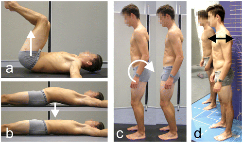
After 12 weeks, the posture test and the examination of the isometric maximum strength were repeated for all 54 participants.
2.4. Statistics and examination of pre-intervention effects
In addition to the descriptive procedures, such as mean values and standard deviations, 95% confidence intervals were calculated.
To assess the performance development of the two intervention groups STR + SENSO and STR as well as of the control group CON, possible pre-test group differences were checked using one-way ANOVA for the variables age, weight, height, pelvic angle PA, and the maximum isometric torque of abdominal muscles (RA), gluteus maximus (GM), and hamstrings (HA). The conditions were controlled applying the Kolmogorov-Smirnov test including the Lilliefors correction for normal distribution and the Levene test for variance homogeneity.
Subsequently, to check possible intervention effects, two-way mixed ANOVA were calculated. Post-hoc analyses were carried out using the Fisher (LSD) test.
The significance level was set to p < 0.05 and statistical relevance was determined by Eta2 (η2). A small effect exists with η2 ≥ 0.01, η2 ≥ 0.06 shows an average effect, and η2 ≥ 0.14 represents a major effect (Cohen, Citation1988).
3. Results
No significant differences were identified between the assigned groups prior to intervention, for age (F = 0.2; p = 0.82), pelvic angle PA (F = 0.07; p = 0.94), relative torque of HA (F = 0.002; p = 0.99), RA (F = 0.2; p = 0.82), and GM (F = 0.56; p = 0.58). Therefore, the test persons are considered to have been matched successfully.
3.1. Intervention effect “pelvic angle PA”
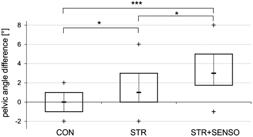
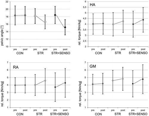
The factor measurement time became significant with major effect size (F = 42.85; p < 0.0001; η2 = 0.46; ), the interaction of measurement time × group became significant too with major effect size (F = 15.65; p < 0.0001; η2 = 0.38). Significant group differences were identified post hoc for the PA differences caused by the treatment between CON (PA reduction: −0.1° ± 1.2°) and STR (PA reduction: −1.6° ± 1.8°) with p = 0.013, as well as between STR and STR + SENSO (PA reduction: −3.3° ± 2.2°) with p = 0.042, and between CON and STR + SENSO with p < 0.0001 ().
3.2. Intervention effect “strength of the hamstrings HA”
The factor measurement time became significant with major effect size (F = 18.68; p < 0.001; η2 = 0.27). A significant interaction effect of time × group was found (F = 3.67; p = 0.029; η2 = 0.13). The pure main effect group was not significant (F = 0.11; p = 0.89). The two intervention groups showed larger improvements than CON (increase 1%), and STR + SENSO (increase 9.6%) in turn showed larger growths than STR (increase 6.6%, ).
3.3. Intervention effect “strength of the rectus abdominis RA”
The factor measurement time became significant with major effect size (F = 18.64; p < 0.0001; η2 = 0.27). Also, a significant interaction effect of measurement time and group (F = 4.14; p = 0.018; η2 = 0.14) was identified. The pure main effect group was not significant (F = 0.24; p = 0.79). Both intervention groups improved almost identically by 13.8% (STR) and 15.0% (STR + SENSO), respectively, while CON did not exhibit any improvements ().
3.4. Intervention effect “strength of the gluteus maximus GM”
The relative torque of GM did not improve between pre- and post-test (F = 3.85; p = 0.06). There was also no significant interaction effect of time × group (F = 1.04; p = 0.36), and neither between the groups (F = 0.66; p = 0.52).
4. Discussion
4.1. Improvement of muscular strength
In both intervention groups, resistance training resulted in a significant improvement of the isometric torque of HA and RA with average and high effect size, respectively. The increase of torque in the range of 9–14% for HA and RA corresponds to the results of other studies that observed similar effects in adolescents within the framework of strength training (Matos & Winsley, Citation2007). Klee (Citation1994) already showed that increasing the maximum strength of the muscles reducing anterior pelvic tilt could result in reduced anteversion in a resting position. He examined the pelvic tilt in adolescents during a 10-week strength and stretch training, and was able to measure an average increase in retroversion of the pelvis of 2.2°.
The increase in strength in the GM was low and did not reach significance. Klee supposed that the lower back and gluteal muscles can only be exercised to a lower degree in the age group examined in a time period of 12 weeks (Klee, Citation1994). Therefore, the relative short time period of the exercises could explain the low increase in strength (López-Miñarro et al., Citation2012). The question remains as to what extent increased maximum strength may influence muscular balance in a relaxed position. The fact that strength training led to an improved pelvic position even in a relaxed position–i.e. without voluntary muscle activation—can be explained by the positive effect of strength training on the stability of sensorimotor coordination (Carroll, Benjamin, Stephan, & Carson, Citation2001) and improvement of neuromotor control (Wong & Ng, Citation2010). An increase in resting length, or rather in range of motion, especially of the hip flexors (M. iliopsoas), might be a further reason (Bridger et al., Citation1992; López-Miñarro et al., Citation2012).
It is interesting to state that the pelvic tilt angles in the training group that performed additional sensorimotor exercises for body perception were significantly better (in the sense of reduced pelvic tilt) than those in the strength training group. Tsaklis, Karlsson, Grooten, and Äng (Citation2008) were able to show that a combination of back-muscle-strengthening and sensorimotor training resulted in improved postural steadiness, which they objectified via the center of pressure variation by means of posturography. Our results additionally show “macroscopic” visible effects on posture.
The positioning of the pelvis in the sagittal plane is determined by the interaction of skeletal, ligamentous, and muscular factors. The capacity of the postural musculature is influenced not only by strength-endurance components, but also by central-nervous activation in the sense of the muscular tone at rest that also affects functional muscular length (Sousa, Silva, & Tavares, Citation2012). It is impossible to say exactly whether the tone of the muscles responsible for the actual positioning of the pelvis is associated with the maximum isometric strength or torque. However, the results measured do suggest just this. Our examinations, as well as previous work (Klee, Citation1994), suggest a connection between maximal isometric muscle strength and position of the pelvis. The improvement of pelvic tilt can therefore be interpreted as the result of changed neuromuscular activation due to the effects of strength and stretch training (Gabriel, Kamen, & Frost, Citation2006).
4.2. Sensorimotor aspects
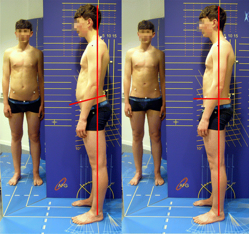
Interestingly, the test persons who had performed additional sensorimotor training showed a significant improved anterior pelvic angle after the end of the intervention. For the relaxed (habitual) posture usually only minimal muscle activity is measurable (Sousa et al., Citation2012). Therefore, the question arises as to how this positive influence of sensorimotor training can be explained. Numerous studies have examined the effect of sensorimotor training on posture and balance parameters (Bruhn, Kullmann, & Gollhofer, Citation2004; Freyler, Weltin, Gollhofer, & Ritzmann, Citation2014; Hwang, Bae, Do Kim, & Kim, Citation2013). Sensorimotor exercises usually include primarily unspecific exercises during which the test person was standing on a flexible platform (unstable spring platforms, wobble boards, etc.), or postural reactions were elicited through targeted disturbances of the equilibrium (Zemková, Citation2009). In contrast to these therapeutic approaches, we trained the perception of the position of the pelvis and the conscious correction of this position. Many children and adolescents are not able to actively execute retroversion of the pelvis (i.e. reduction of pelvic tilt) in a controlled manner, they have to learn this. Training this develops an idea of posture and movement of the pelvis (). Through sensorimotor training, the subjects learn how to adjust muscle tension in order to generate a specific movement or position (Hwang et al., Citation2013) and thus improve sensor integration (Peterka, Citation2002). The present results can be explained by the fact that improved posture and movement perception can reinforce the effects that would have been created by pure strength training. This finding is in accordance with a study by Hwang et al. (Citation2013) who were able to show that body perception exercises improved anticipatory postural adjustments. Tsaklis et al. (Citation2008) see increased sensitivity of feedback pathways from proprioceptive sensory input as a reason for this.
Figure 7. Voluntary reduction of pelvic tilt by a 16 year old subject after the exercise program (STR + SENSO).
Note: The decrease in pelvic angle that results in a reduction of the lumbar lordosis and the improved alignment of the body in relation to the perpendicular.
It can be ruled out that the greater effects measured in the STR + SENSO group are induced by the sensorimotor exercises because a difference in muscle strength between STR and STR + SENSO was not verified for any of the muscle groups trained and tested. The STR and the STR + SENSO training groups increased their maximum strength values equally. Therefore, any additional effect can be attributed to the additional sensorimotor exercises.
Moreover, there seems to be no explicit interaction between strength and balance parameters. Kaminski, Buckley, Powers, Hubbard, and Ortiz (Citation2003) were able to prove that balance training did not influence muscle torque. The same result was found by Zech et al. (Citation2010), considering balance training as an effective intervention to improve static and dynamic balance, but with no effects for back extensor muscle strength. Vice-versa, Kollmitzer, Ebenbichler, Sabo, Kerschan, and Bochdansky (Citation2000) elaborated that back extensor strengthening led to decreased postural stability.
While strength training improves mainly intramuscular coordination and since the effects observed mainly occur in the area of efferent pathways (Bruhn et al., Citation2004), sensorimotor exercises presumably improve mainly the processing of sensory input. This could result in improved tone regulation of the muscle groups involved and could have enhanced intermuscular coordination.
According to our knowledge, this study is the first to show that sensorimotor physical therapy exercises to improve body perception in the pelvic region performed before strength and stretch training led to an improvement of the effectiveness of the training. This finding could supplement existing fitness and physical therapy intervention programs (Kim, Cho, Park, & Yang, Citation2015; Yoo, Citation2013) for posture improvement in children and adolescents.
4.3. Limitations
The study was subject to various limitations. While the subjects did exhibit an above-normal pelvic tilt, they were symptom-free. Further research could identify the extent to which functional sensorimotor training affects patients with lower back pain. At this point, it is impossible to make any statements on long-term effects on the pelvic position.
4.4. Conclusion
Physical fitness training programs including sensorimotor exercises to improve body perception in the pelvic area improve the pelvic tilt in adolescents more than pure strength and stretching exercises. Therefore, it is recommended that sensorimotor exercises become a permanent part of physical training programs to preventively treat poor posture caused by increased pelvic tilt.
Declaration
The study protocol was approved by the Ethics Committee of the Saarland University in Saarbrücken, Germany (ref. 15–6).
Funding
The authors received no direct funding for this research.
Acknowledgment
The authors wish to thank Monika Schutz for translating the manuscript.
Additional information
Notes on contributors
Oliver Ludwig
Dr Oliver Ludwig is a human biologist and scientific head of the Kid-Check working group at Saarland University. The Kid-Check working group consists of orthopedists, pediatricians, bioscientists, sports scientists, and physical therapists researching posture weakness in children and adolescents. One objective is the monitoring of interventions, such as special training programs, that are supposed to effectively improve poor posture. The research reported in this paper relates to this project. The scientists involved have also developed measurement methods to analyze body posture in a reproducible way and explore interrelationships between posture and movement. More than 1,500 children and adolescents have been examined since the year 2000.
References
- Alvim, F. C., Peixoto, J. G., Vicente, E. J., Chagas, P. S., & Fonseca, D. S. (2010). Influences of the extensor portion of the gluteus maximus muscle on pelvic tilt before and after the performance of a fatigue protocol. Revista Brasileira de Fisioterapia, 14, 206–213.
- Bogdanović, Z., & Marković, Ž. (2010). Presence of lordotic poor posture resulted by absence of sport in primary school children. Acta Kinesiologica, 4, 63–66.
- Bridger, R. S., Orkin, D., & Henneberg, M. (1992). A quantitative investigation of lumbar and pelvic postures in standing and sitting: Interrelationships with body position and hip muscle length. International Journal of Industrial Ergonomics, 9, 235–244. doi:10.1016/0169-8141(92)90017-T
- Bruhn, S., Kullmann, N., & Gollhofer, A. (2004). The effects of a sensorimotor training and a strength training on postural stabilisation, maximum isometric contraction and jump performance. International Journal of Sports Medicine, 25, 56–60. doi:10.1055/s-2003-45228
- Bruhn, S., Kullmann, N., & Gollhofer, A. (2006). Combinatory effects of high-intensity-strength training and sensorimotor training on muscle strength. International Journal of Sports Medicine, 27, 401–406.10.1055/s-2005-865750
- Buchtelová, E., Tichy, M., & Vaniková, K. (2013). Influence of muscular imbalances on pelvic position and lumbar lordosis: A theoretical basis. Journal of Nursing, Social Studies, Public Health and Rehabilitation, 1–2, 25–36.
- Carroll, T. J., Benjamin, B., Stephan, R., & Carson, R. G. (2001). Resistance training enhances the stability of sensorimotor coordination. Proceedings of the Royal Society B: Biological Sciences, 268, 221–227.10.1098/rspb.2000.1356
- Chaléat-Valayer, E., Mac-Thiong, J.-M., Paquet, J., Berthonnaud, E., Siani, F., & Roussouly, P. (2011). Sagittal spino-pelvic alignment in chronic low back pain. European Spine Journal, 20, 634–640. doi:10.1007/s00586-011-1931-2
- Cohen, J. (1988). Statistical power analysis for the behavioral sciences (2nd ed.). Hillsdale, NJ: Lawrence Erlbaum Associates.
- Dolphens, M., Cagnie, B., & Coorevits, P. (2012). Sagittal standing posture and its association with spinal pain. Spine, 37, 1657–1666.10.1097/BRS.0b013e3182408053
- Freyler, K., Weltin, E., Gollhofer, A., & Ritzmann, R. (2014). Improved postural control in response to a 4-week balance training with partially unloaded bodyweight. Gait and Posture, 40, 291–296. doi:10.1016/j.gaitpost.2014.04.186
- Gabriel, D. A., Kamen, G., & Frost, G. (2006). Neural adaptations to resistive exercise. Sports Medicine, 36, 133–149.10.2165/00007256-200636020-00004
- Gajdosik, R., Simpson, R., Smith, R., & DonTigny, R. L. (1985). Pelvic tilt. Intratester reliability of measuring the standing position and range of motion. Physical Therapy, 65, 169–174.
- Gh, M. E., Alilou, A., Ghafurinia, S., & Fereydounnia, S. (2012). Prevalence of faulty posture in children and youth from a rural region in Iran. Biomedical Human Kinetics, 4, 121–126.
- Hwang, J. A., Bae, S. H., Do Kim, G., & Kim, K. Y. (2013). The effects of sensorimotor training on anticipatory postural adjustment of the trunk in chronic low back pain patients. Journal of Physical Therapy Science, 25, 1189–1192. doi:10.1589/jpts.25.1189
- Jentzsch, T., Geiger, J., Bouaicha, S., Slankamenac, K., Nguyen-Kim, T. D., & Werner, C. M. (2013). Increased pelvic incidence may lead to arthritis and sagittal orientation of the facet joints at the lower lumbar spine. BMC Medical Imaging, 13, 556. doi:10.1186/1471-2342-13-34
- Kaminski, T. W., Buckley, B. D., Powers, M. E., Hubbard, T. J., & Ortiz, C. (2003). Effect of strength and proprioception training on eversion to inversion strength ratios in subjects with unilateral functional ankle instability * commentary. British Journal of Sports Medicine, 37, 410–415.10.1136/bjsm.37.5.410
- Kim, D., Cho, M., Park, Y., & Yang, Y. (2015). Effect of an exercise program for posture correction on musculoskeletal pain. Journal of Physical Therapy Science, 27, 1791–1794. doi:10.1589/jpts.27.1791
- Kim, H. J., Chung, S., Kim, S., Shin, H., Lee, J., Kim, S., & Song, M. Y. (2006). Influences of trunk muscles on lumbar lordosis and sacral angle. European Spine Journal, 15, 409–414. doi:10.1007/s00586-005-0976-5
- Klee, A. (1994). Posture, muscular balance and training (Vol. 20). Frankfurt/Main: Verlag Harri Deutsch.
- Kollmitzer, J., Ebenbichler, G. R., Sabo, A., Kerschan, K., & Bochdansky, T. (2000). Effects of back extensor strength training versus balance training on postural control. Medicine and Science in Sports and Exercise, 32, 1770–1776.10.1097/00005768-200010000-00017
- Kopecký, M. (2004). Posture assessment in children of the school age group (7–15 years of age) in the Olomouc Region. Acta Universitatis Palackianae Olomucensis Gymnica, 34, 19–29.
- Laird, R. A., Gilbert, J., Kent, P., & Keating, J. L. (2014). Comparing lumbo-pelvic kinematics in people with and without back pain: A systematic review and meta-analysis. BMC Musculoskeletal Disorders, 15, 242–229. doi:10.1186/1471-2474-15-229
- Le Huec, J. C., Aunoble, S., Philippe, L., & Nicolas, P. (2011). Pelvic parameters: Origin and significance. European Spine Journal, 20, 564–571. doi:10.1007/s00586-011-1940-1
- López-Miñarro, P. A., Muyor, J. M., Belmonte, F., & Alacid, F. (2012). Acute effects of hamstring stretching on sagittal spinal curvatures and pelvic tilt. Journal of Human Kinetics, 31, 69–78. doi:10.2478/v10078-012-0007-7
- Matos, N., & Winsley, R. J. (2007). Trainability of young athletes and overtraining. Journal of Sports Science and Medicine, 6, 353–367.
- McCaskey, M. A., Schuster-Amft, C., Wirth, B., Suica, Z., & de Bruin, E. D. (2014). Effects of proprioceptive exercises on pain and function in chronic neck- and low back pain rehabilitation: A systematic literature review. BMC Musculoskeletal Disorders, 15, 29. doi:10.1186/1471-2474-15-382
- Nguyen, A. D., & Shultz, S. J. (2007). Sex differences in clinical measures of lower extremity alignment. Journal of Orthopaedic and Sports Physical Therapy, 37, 389–398. doi:10.2519/jospt.2007.2487
- Nourbakhsh, M. R., & Arab, A. M. (2002). Relationship between mechanical factors and incidence of low back pain. Journal of Orthopaedic and Sports Physical Therapy, 32, 447–460. doi:10.2519/jospt.2002.32.9.447
- Page, P. (2006). Sensorimotor training: A “global” approach for balance training. Journal of Bodywork and Movement Therapies, 10, 77–84. doi:10.1016/j.jbmt.2005.04.006
- Peterka, R. (2002). Sensorimotor integration in human postural control. Journal of Neurophysiology, 88, 1097–1118.
- Roussouly, P., & Pinheiro-Franco, J. L. (2011). Biomechanical analysis of the spino-pelvic organization and adaptation in pathology. European Spine Journal, 20, 609–618. doi:10.1007/s00586-011-1928-x
- Ryan, E. E., Rossi, M. D., & Lopez, R. (2010). The effects of the contract-relax-antagonist-contract form of proprioceptive neuromuscular facilitation stretching on postural stability. Journal of Strength and Conditioning Research, 24, 1888–1894. doi:10.1519/JSC.0b013e3181ddad9d
- Smith, A., O'Sullivan, P., & Straker, L. (2008). Classification of sagittal thoraco-lumbo-pelvic alignment of the adolescent spine in standing and its relationship to low back pain. Spine, 33, 2101–2107. doi:10.1097/BRS.0b013e31817ec3b0
- Sousa, A. S., Silva, A., & Tavares, J. M. (2012). Biomechanical and neurophysiological mechanisms related to postural control and efficiency of movement: A review. Somatosensory and Motor Research, 29, 131–143. doi:10.3109/08990220.2012.725680
- Tsaklis, P., Karlsson, J., Grooten, W., & Äng, B. (2008). A combined sensorimotor skill and strength training program improves postural steadiness in rhythmic sports athletes. Human Movement, 9, 34–40.
- Wong, Y., & Ng, G. (2010). Resistance training alters the sensorimotor control of vasti muscles. Journal of Electromyography and Kinesiology, 20, 180–184.10.1016/j.jelekin.2009.02.006
- Yoo, W. G. (2013). Effect of individual strengthening exercises for anterior pelvic tilt muscles on back pain, pelvic angle, and lumbar roms of a lbp patient with flat back. Journal of Physical Therapy Science, 25, 1357–1358. doi:10.1589/jpts.25.1357
- Zech, A., Hübscher, M., Vogt, L., Banzer, W., Hänsel, F., & Pfeifer, K. (2010). Balance training for neuromuscular control and performance enhancement: A systematic review. Journal of Athletic Training, 45, 392–403. doi:10.4085/1062-6050-45.4.392
- Zemková, E. (2009). The acute and long-term effect of different sensorimotor exercises on neuromuscular performance. Medicina Sportiva, 13, 67–73.10.2478/v10036-009-0012-7

