
Abstract
Purpose: To evaluate the degree of fibroid infarction after uterine fibroid embolization (UFE) with tris-acryl gelatin microsphere (Embosphere) visualized by contrast-enhanced magnetic resonance imaging (MRI) and evaluation of the clinical outcome measured with symptom and quality of life questionnaire (UFS-QOL) after 3 months follow-up. Materials and Methods: A prospective study included twenty-six pre-menopausal women. UFE with Embosphere of 500–900 μm was performed. Residual contrast enhancement in the total fibroid burden was analyzed and residual contrast enhancement ≤10% was defined as a satisfactory result after embolization. The dominant fibroids and total uterine volumes were assessed by MRI before and at follow-up. The UFS-QOL questionnaire answers were analysed and compared. Results: Twenty-five of 26 patients (96%) had a technically successful UFE. Twenty-four of the patients (92%) had MRI controls 3 months after UFE of which 23 patients had complete or almost complete fibroid burden infarction without residual contrast enhancement of the fibroids of more than 10%. One patient had insufficient fibroid burden infarction. Twenty-three of the patients had UFS-QOL analyses of which 96% had significant clinical improvement. Conclusion: UFE with Embosphere of 500–900 μm gave satisfactory MRI results with residual fibroid contrast enhancement ≤10% and good clinical effect in 96% of patients.
Public Interest Statement
The uterine fibroid is very frequent female benign tumour with impact on quality of life in a symptomatic patients. The mains symptoms are bleedings, pressure symptoms and pain. I some cases the fibroid cause infertility. The surgical treatment was the standard treatment, but after the well-conducted randomised study, the majority of the Europea and North American Gynaecological society include minimally invasive treatment such as uterine artery embolization (UAE) as a treatment option. With UAE the quality of life and symptoms control are achieved in a majority of the patients. However, the reintervention rate is more frequent compared with the surgical hysterectomy. Compared with the another uterus sparing surgical modality re-intervention is similar.
The unanswered question is safety of UAE in a patients who want to be pregnant. There is no strong evidence to support either the myomectomy or UAE in this group of patients and more evidence is needed.
Competing Interests
The authors declare no competing interest.
1. Introduction
Uterine fibroid embolization (UFE) is a well-established treatment of symptomatic fibroids with clinical efficacy comparable with traditional surgical treatment (Gupta, Sinha, Lumsden, & Hickey, Citation2014). Since introduction there has been a lot of investigations of fibroid characteristics, imaging of the fibroids, technical details during interventions and investigation of the available embolic agents regarding clinical and imaging outcome (Katsumori, Kasahara, Kin, & Nozaki, Citation2008; Spies, Citation2013). The first generation of microparticles is non-spherical polyvinyl alcohol particles (ns-PVA, Boston Scientific, Natick, MA, USA and Ivalon, Cook Medical, Bloomington, IN, USA) which showed favourable results but the non-spherical shape had some disadvantages and following the calibrated spheres has been developed. Currently, there are five types of microspheres available, tris-acryl gelatin microsphere (Embosphere, Merit Medical, Paris, France), Acrylamido polyvinyl alcohol microsphere (Bead Block, Biocompatibles, Farnham UK), polymer Polyzene-F (Embozene, CeloNova, Bioscience San Antonio, Texas, USA), Polyethylene glycol microsphere (HydroPearl, Terumo Europe, Leuven, Belgium) and spherical polyvinyl alcohol microsphere (Contour SE, Boston Scientific, Natick, MA, USA). The clinical and imaging outcomes after use of spherical polyvinyl alcohol microspheres (s-PVA) and Embosphere have been studied extensively showing that Embospheres achieved better results (Rasuli et al., Citation2008; Siskin et al., Citation2008; Spies et al., Citation2005). The potential difference between Embosphere and Bead Block has not been studied extensively, and there is still no evidence in favour of any of these microspheres (Das, Champaneria, Daniels, & Belli, Citation2014).
In the upstart of UFE in 1999 at our institution we used ns-PVA and switched after introduction of spherical microspheres on the market to s-PVA, but the results and achievement with this type of embolic agent were not optimal (Duvnjak, Ravn, Green, & Andersen, Citation2016). During the latest few years, we have used Bead Block as an embolic agent for UFE but demonstrated insufficient fibroid infarction in 27% of the patients (Duvnjak, Ravn, Green, & Andersen, Citation2017). Therefore a prospective study was initiated to investigate completeness of fibroid infarction and residual contrast enhancement of the total fibroid burden after UFE using the Embosphere of 500–900 μm in size as embolic agent.
The working hypothesis was that Embosphere might be a more potent embolic agent than the others and can give more complete embolization in a majority of the patients.
Primary aim of the study was to evaluate the rate of fibroid infarction and the residual total fibroid burden after UFE with Embosphere demonstrated by contrast-enhanced magnetic resonance imaging (MRI). Secondary aim was to investigate the clinical outcome after three months follow-up using a validated uterine fibroid symptom and quality of life (UFS-QOL) questionnaire (Spies et al., Citation2002).
2. Material and methods
Informed consent was waived for all participants, and the regional ethic committee approved the study (S-20120138). Twenty-six patients with symptomatic fibroids entered the study from February 2015 to May 2016. The baseline non-contrast- and contrast enhanced MRI before UFE were performed in all cases. The follow-up protocol included three months post UFE MRI control. All patients were asked to answer the UFS-QOL questionnaire at baseline and at follow-up three months after UFE.
2.1. Study design
All consecutive symptomatic patients with menorrhagia, metrorrhagia, bulk-related symptoms or a combination of these symptoms who accepted to participate were included in the study. The gynaecologist examined all patients and confirmed the diagnosis of symptomatic fibroids and excluded other diseases. The residual contrast enhancement of the fibroids and the uterine and dominant fibroid volumes was analysed by MRI and compared with baseline data at three months MRI control. The official Danish translation of the UFS-QOL questionnaire was provided by the authors and permission to use the translated questionnaire subsequently granted by the Society of Interventional Radiology. Exclusion criteria were: asymptomatic fibroids, active pelvic infection, malignancy, pregnancy, age <18 years and >52 years, non-contrast enhancing of the fibroids at the baseline examination and adenomyosis.
2.2. Intervention
All procedures were performed under local anaesthesia. UFE was performed by the same IR specialist, in all but one case using transfemoral percutaneous access and in one case by left trans-brachial access due to obesity. The patients had a self-controlled morphine analgesia pump. Peroral pain drugs and antiemetic drugs were prescribed when needed. After selective bilateral catheterization of the internal iliac arteries, “microcatheter” (Direxion HI-FLO, inner lumen 0,027″ Boston Scientific, Natick,MA,USA) “microcatheter” were placed in a transversal part of the uterine artery. The embolization with Embosphere microspheres was performed until “near” flow-stop defined as sluggish flow through the horizontal part of the main uterine artery and flow stop in the peri fibroid arteries on the both sides. The patients were embolized with 500–700 μm microspheres and if the desired end-point was not achieved after 6 ml of these microspheres embolization were followed by 700–900 μm microspheres till near flow stop. The achievement of the end-point was confirmed after five minutes waiting by flushing contrast in all cases. In case of incomplete flow-stop, supplementary embolization was performed. Finally, abdominal aortography was performed to disclose possible fibroid supply from the ovarian arteries. At the discretion of the patients, it was decided whether or not to perform supplementary embolization of the ovarian artery.
2.3. MRI analyses
MRI using phased array torso coil was performed with 1.5 Tesla MR Systems, (Philips Medical System, the Netherlands). The standard MR fibroid protocol consists of non-contrast T1, and T2 weighted axial and sagittal as well as fat saturated post contrast T1 trans-axial gradient recalled echo and turbo spin echo image sequences (Kirby, Burrows, Haider, Maizlin, & Midia, Citation2011). The largest dominant fibroid volume and total uterine volume using the ellipsoid formula (length × height × width × 0, 52), the number of fibroids and dominant fibroid localisation were analysed and recorded. The fibroid burden infarction after UFE and residual fibroid burden contrast enhancement were visually estimated by contrast-enhanced MRI at follow-up after three months. Results were stratified into three categories: category 1: 100% fibroid infarction = no residual enhancement (Figure (A)–(B)), category 2: 99–90% fibroid infarction with residual fibroid contrast enhancement less than 10% (Figure (A)–(B)), and category 3: less than 90% fibroid infarction with residual contrast fibroid enhancement more than 10% (Figure (A)–(B)). The first two categories were defined as successful outcomes and category three as an unsuccessful outcome. Dominant fibroid infarction, as well as complete fibroid burden infarction, were analysed as described above. The fibroid and uterine volume changes were reviewed accordingly at follow-up images and compared with baseline measurement. Two radiologists, one with over 7-years’ experience in pelvic MRI imaging and the other with 5 years experience reviewed the images in consensus in a non-blinded fashion.
Figure 1. (A) Presents contrast-enhanced MRI of the fibroid prior to UFE. (B) Presents the same fibroid after UFE with complete fibroid infarction (100%) without residual contrast enhancement. Category 1.
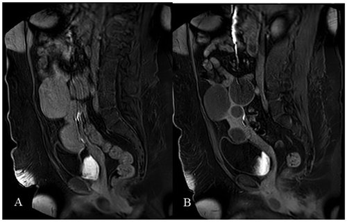
Figure 2. (A) Presents contrast-enhanced MRI of the fibroid prior to UFE. (B) Presents the same fibroid after UFE with almost complete fibroid infarction (90–99%) and residual contrast enhancement below 10%. Note small irregularity on the base of embolized fibroid. Category 2.
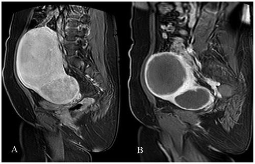
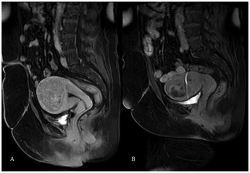
Figure 3. (A) Presents contrast-enhanced MRI of the fibroid prior to UFE. (B) present the same fibroid after UFE with incomplete fibroid infarction (<90%) and with residual contrast enhancement over 10%. The central part of the fibroid is with residual contrast enhancement. Category 3.
2.4. UFS-QOL questionnaire analyse
The UFS-QOL questionnaire consists of an 8-item symptom severity scale and 29-item health-related quality of life (HRQOL) comprising six domains: Concern, activities, energy/mood, control, self-consciousness, and sexual function. The Likert 5-point scale, ranging from “not at all” to “a very great deal” for symptom severity score and a range from “none of the time” to “all of the time” for the HRQOL score was used for questionnaire analysis. Both scores were summed and transformed into a 0–100 point scale (Spies et al., Citation2002). High symptom severity score indicates severe symptoms and a high HRQOL score indicates a good quality of life.
2.5. Statistical analyze
Continuous variables are expressed as mean (±standard deviation-SD) or median, as appropriate. Nominal data are given as absolute numbers and percentages. Numerical variables were compared using two-tailed Student’s t-test. Wilcoxon paired sample test was used to compare results of the UFS-QOL questionnaire score using the SSPS software package (Statistics 21 IBM Corporation NY, USA). Statistical significance was set at p < 0. 05
3. Results
In the period February 2015 until May 2016, 31 patients were referred for UFE. Twenty-six patients were included in the study while four patients desisted to participate and one patient was excluded because of high age.
3.1. Baseline characteristics
The mean age in the entire group was 44.5 year (range: 32–52). The patients’ dominant symptoms and MRI fibroid characteristics are presented in Table .
Table 1. Patients symptoms and MRI characteristics
3.2. UFE intervention
Twenty-five patients (96%) had a technically successful UFE performed with complete flow-stop in both uterine arteries and no associated peri-procedural complications. The end-point of near complete stasis of flow was achieved in all cases. In one patient (4%) the left uterine artery was too small and hypoplastic to allow safe particle administration without the risk of reflux and following non-target embolization. There was one case with additional left ovarian artery fibroid vascular supply left untreated due to the patient’s wish. There were no cases which required additional embolization after five minutes waiting. Prophylactic antibiotics were not administrated in any case according to our treatment protocol. The average volume of microspheres used per patient was 9.1 ml (range: 6–16 ml).
3.3. MRI follow-up
Twenty-four patients (92%) completed three months follow-up with MRI. One patient did not complete three months control and withdrew from the study and another patient underwent hysterectomy two months after UFE. Twenty-three of the 24 patients, (96%) had complete fibroid burden infarction and/or residual contrast enhancement of the fibroid burden of less than 10% at three months MRI control (category 1 and/or 2). One of the 24 patients, (4%) had insufficient dominant fibroid infarction with residual contrast enhancement of more than 10% (category 3). The left ovarian artery was left patent in this patient which might be responsible for the residual contrast enhancement. The other patient with a hypoplastic left uterine artery which was not embolized had on follow-up MRI no residual enhancement in the fibroids (Figure (A)–(C)). The median dominant fibroid volume reduction was presented with a statistically significant reduction of 49% after three months follow-up (p < 0.01). The median uterine volume reduction was 47% which was a significant reduction compared with baseline (p < 0.007).
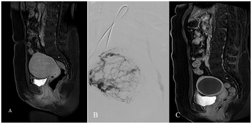
Figure 4. (A) Presents fibroid prior to UFE. The left uterine artery was not embolized due to small calibre. (B) Presents almost complete fibroid vascularization from the right uterine artery. (C) Presents control contrast enhancing MRI with complete fibroid infarction despite the left uterine artery was not embolized.
3.4. UFS-QOL questionnaire analysis-clinical outcome
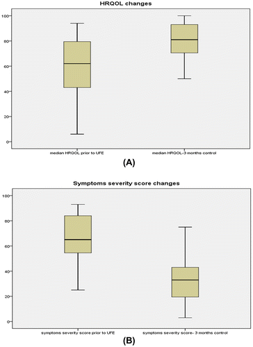
Twenty-three patients completed the UFS-QOL questionnaires including baseline and three months follow-up. The median HRQOL score prior to UFE was 62 (range: 6–94) and had improved significantly after three months to 81 (p < 0.001). The median symptom severity score prior to UFE was 65 (range: 25–93) and had been significantly decreased after three months to 33 (p < 0.001). In Figure the median HRQL score and symptom severity score changes are graphically presented. Only one patient (4%) had an increased symptom severity score. Two patients (9%) had unchanged HRQOL score, but these patients at the same time had significant decreasing of symptom severity score.
Figure 5. The median changes in the HRQOL and symptoms severity score. (A) Significant increasing in HRQOL after UFE (p < 0.001). (B) Significant decreasing of symptoms severity score (p < 0.001).
Complications were classified according to the SIR classification (Hovsepian et al., Citation2009). One patient (5%) underwent hysterectomy two months after UFE due to symptom persistence and hospital admission >48 h, classified as SIR complication D. Two patients (9%) had non-bacterial endometritis treated with antibiotics without further problems. They required admission < 48 h and have been classified as SIR complication C due to pain and prolonged discharge, one patient had an extended hospital admission but <48 h, which was classified as SIR complication C also. Three patients experienced clinical “silent” fibroid expulsion requiring no further treatment.
4. Discussion
UFE has an established role in the treatment of symptomatic fibroids (Hehenkamp, Volkers, Birnie, Reekers, & Ankum, Citation2008; Manyonda et al., Citation2012; The REST Investigators, Citation2007). During the last two decades, technical and imaging improvements have contributed to the mastering of the intervention. There are not many unknown remaining issues during UFE, especially from the technical point of view. After Spies in 2005 (Spies, Citation2009) questioned what type of evidence we need before adopting a new embolic agent, the level of evidence has been increasing (Golzarian et al., Citation2006; Spies, Cornell, Worthington-Kirsch, Lipman, & Benenati, Citation2007). Further, MRI control is essential to ensure quality after UFE and is the most commonly used modality for follow-up (Katsumori et al., Citation2008; Kroencke, Scheurig, Poellinger, Gronewold, & Hamm, Citation2010). The still not so clear issue is a behaviour of the different embolic agents and availability to obtain complete fibroid infarction, or precise some of them. Das et al. (Citation2014) in a meta-analysis emphasised that still, we do not have strong evidence comparing Embosphere with Bead Block microspheres. The new embolic agent like Embozene and HydroPearl are not included in the meta-analysis as well.
The fibroid infarction degree has a strong correlation with especially long-term clinical successful outcome where patients with fibroid infarction ≥90% have better clinical results comparing with patients with fibroid infarction below 90% (Katsumori et al., Citation2008; Koesters, Powerski, Froeling, Kroencke, & Scheurig-Muenkler, Citation2012; Worthington-Kirsch, Siskin, & Hegener, Citation2009). Katsumori et al. (Citation2008) showed at five year follow up that cumulative rate of symptom control was 93% for patients with complete fibroid infarction compared regarding symptom control in only 60% of the patients with fibroid infarction below 90%. Further, Koesters et al. (Citation2012) reported that among 22 patients with complete fibroid infarction, 20 patients had no symptom recurrence after eight years follow-up, in contrast with six patients with fibroid infarction below 90% who all had symptom recurrence. Gabriel-Cox, Jacobson, Armstrong, Hung, and Learman (Citation2007) concluded that incomplete fibroid infarction is the most likely cause of recurrence of symptoms. They compared patients with unintended unilateral UFE who had a re-intervention rate of 39.2% with patients with bilateral UFE which had a re-intervention rate 18.5%. In conclusion, the majority of the authors suggest that fibroid infarction should be ≥90%, but some authors suggest ≥80% of fibroid infarction as sufficient result (Smeets et al., Citation2010).
The present study showed that 96% of the patients obtained complete or almost complete fibroid infarction after UFE with the use of Embosphere 500–900 μm in size. Only one patient had insufficient MRI result with a residual fibroid burden contrast enhancement above 10%, probably due to additional fibroid vascular supply from the ovarian artery. In a recently published study showed inferior performance of Bead Block particles compared with Embosphere particles which were used in the present study (Duvnjak et al., Citation2017). The patients’ demography, symptoms, and embolization technique and endpoints were comparable in the two studies which allow us to draw this conclusion. Spies et al. (Citation2005) achieved similar high infarction rate in their study using Embosphere and reported residual contrast enhancement in 5% of the patients. The same study showed residual contrast enhancement in 44.7% of the patients embolized with s-PVA.
The complications rate in the present study, especially fibroid expulsion, was a little higher compared with other studies (Golzarian et al., Citation2006; Hehenkamp et al., Citation2008; Manyonda et al., Citation2012; Worthington-Kirsch et al., Citation2009) and one of the explanations could be submucosal and partial submucosal fibroid localisation in two cases. Endometritis is the other complication and can be explained with extensive embolization and following ischemia of the surrounding myometrial tissue. However, absent of major complications and clinical improvement are in line with other published results (Andersen et al., Citation2001; Spies, Citation2013).
The main limitations of this study are short-term follow-up and a relatively small number of patients. The other limitations are non-blinded MRI review and not a randomised comparison with the other embolic agents.
In conclusion, Embosphere (500–900 μm) achieved in 96% of the patients complete or almost complete fibroid burden infarction and present the best results until now in our praxis. Embosphere seems to be the most powerful microspheres for use in UFE today. The newer embolic agents should be examined in a prospective randomised study comparing with Embosphere before wider clinical use.
Funding
The authors received no direct funding for this research.
Additional information
Notes on contributors
S. Duvnjak
S. Duvnjak is Interventional radiologist and together with another colleagues perform variety of endovascular interventions, including embolization, EVAR, TEVAR and peripheral endovascular interventions. Odense University Hospital, Department of Radiology is leading hospital in Denmark in endovascular medicine including the research and scientific works.
References
- Andersen, P. E., Lund, N., Justesen, P., Munk, T., Elle, B., & Floridon, C. (2001). Uterine artery embolization of symptomatic uterine fibroids: Initial success and short-term results. Acta Radiologica, 234, 238.
- Das, R., Champaneria, R., Daniels, J. P., & Belli, A. M. (2014). Comparison of embolic agents used in uterine artery embolisation: A systematic review and meta-analysis. CardioVascular and Interventional Radiology, 37, 1179–1190.10.1007/s00270-013-0790-0
- Duvnjak, S., Ravn, P., Green, A., & Andersen, P. E. (2016). Clinical long-term outcome and reinterventional rate after uterine fibroid embolization with nonspherical versus spherical polyvinyl alcohol particles. CardioVascular and Interventional Radiology, 39, 204–209.10.1007/s00270-015-1157-5
- Duvnjak, S., Ravn, P., Green, A., & Andersen, P. E. (2017). Uterine fibroid embolization with Acrylamido polyvinyl microspheres: Prospective 12 months clinical and MRI follow-up study. Acta Radiologica, 58, 952–958. Epub 2016 Nov 21. doi:10.1177/0284185116679458.
- Gabriel-Cox, K., Jacobson G. F., Armstrong M. A., Hung Y. Y., Learman L. A. (2007). Prediction of hysterectomy after uterine artery embolization for leiomyoma. American Journal of Obstetrics and Gynecology, 196, 588.e1–588.e6.10.1016/j.ajog.2007.03.014
- Golzarian, J., Lang, E., Hovsepian, D., Kroncke, T., Lampmann, L., Lohle, P., … Spies, J. (2006). Higher rate of partial devascularization and clinical failure after uterine artery embolization for fibroids with spherical polyvinyl alcohol. CardioVascular and Interventional Radiology, 29, 1–3.10.1007/s00270-005-0243-5
- Gupta, J. K., Sinha, A., Lumsden, M. A., & Hickey, M. (2014). Uterine artery embolization for symptomatic uterine fibroids. The Cochrane Database of Systematic Reviews, 26, 12.
- Hehenkamp, W. J., Volkers, N. A., Birnie, E., Reekers, J. A., & Ankum, W. M. (2008). Symptomatic uterine fibroids: Treatment with uterine artery embolization or hysterectomy—Results from the randomized clinical embolisation versus hysterectomy (EMMY) trial. Radiology, 246, 823–832.10.1148/radiol.2463070260
- Hovsepian, D. M., Siskin, G. P., Bonn, J., Cardella, J. F., Lapmann, LE, Clark, T. W., … Schwartzberg, M. S. (2009). Quality improvement guidelines for uterine artery embolization for symptomatic leiomyomata. Journal of Vascular and Interventional Radiology, 20, S193–S199.10.1016/j.jvir.2009.04.006
- Katsumori, T., Kasahara, T., Kin, Y., & Nozaki, T. (2008). Infarction of uterine fibroids after embolization: Relationship between postprocedural enhanced MRI findings and long-term clinical outcomes. CardioVascular and Interventional Radiology, 31, 66–72.10.1007/s00270-007-9187-2
- Kirby, J. M., Burrows, D., Haider, E., Maizlin, Z., & Midia, M. (2011). Utility of MRI before and after uterine fibroid embolization: Why to do it and what to look for. CardioVascular and Interventional Radiology, 34, 705–716.10.1007/s00270-010-0029-2
- Koesters, C., Powerski, M. J., Froeling, V., Kroencke, T. J., & Scheurig-Muenkler, C. (2012). Uterine artery embolization in single symptomatic leiomyoma: Do anatomical imaging criteria predict clinical presentation and long-term outcome? Acta Radiologica, 55, 441–449.
- Kroencke, T. J., Scheurig, C., Poellinger, A., Gronewold, M., & Hamm, B. (2010). Uterine artery embolization for leiomyomas: Percentage of infarction predicts clinical outcome. Radiology, 255, 834–841.10.1148/radiol.10090977
- Manyonda, I. T., Bratby, M., Horst, J. S., Banu, N., Gorti, M., & Belli, A. M. (2012). Uterine artery embolization versus myomectomy: Impact on quality of life—results of the FUME (fibroids of the uterus: myomectomy versus embolization) trial. CardioVascular and Interventional Radiology, 35, 530–536.10.1007/s00270-011-0228-5
- Rasuli, P., Hammond, I., Al-Mutairi, B., French, G. J., Aquino, J., Hadziomerovic, A., … Jolly, E. E. (2008). Spherical versus conventional polyvinyl alcohol particles for uterine artery embolization. Journal of Vascular and Interventional Radiology, 19, 42–46.10.1016/j.jvir.2007.08.016
- Siskin, G. P., Beck, A., Schuster, M., Mandato, K., Englander, M., & Herr, A. (2008). Leiomyoma infarction after uterine artery embolization: A prospective randomized study comparing tris-acryl gelatin microspheres versus polyvinyl alcohol microspheres. Journal of Vascular and Interventional Radiology, 19, 58–65.10.1016/j.jvir.2007.08.034
- Smeets, A. J., Nijenhuis, R. J., van Rooij, W. J., Weimar, E. A., Boekkooi, P. F., Lampmann, L. E., & Lohle, P. N. (2010). Uterine artery embolization in patients with a large fibroid burden: Long-term clinical and MR follow-up. CardioVascular and Interventional Radiology, 33, 943–948.10.1007/s00270-009-9793-2
- Spies, J. B. (2009). What evidence should we demand before accepting a new embolic material for uterine artery embolization? Journal of Vascular and Interventional Radiology, 20, 567–570.
- Spies, J. B. (2013). Current evidence on uterine embolization for fibroids. Seminars in Interventional Radiology, 30, 340–346.
- Spies, J. B., Allison, S., Flick, P., Cramp, M., Bruno, J., Jha, R. C., & Ascher, S. A. (2005). Spherical polyvinyl alcohol versus tris-acryl gelatin microspheres for uterine artery embolization for leiomyomas: Results of a limited randomized comparative study. Journal of Vascular and Interventional Radiology, 16, 1431–1437.10.1097/01.RVI.0000179793.69590.1A
- Spies, J. B., Cornell, C., Worthington-Kirsch, R., Lipman, J. C., & Benenati, J. F. (2007). Long-term outcome from uterine fibroid embolization with tris-acryl gelatin microspheres: Results of a multicenter study. Journal of Vascular and Interventional Radiology, 18, 203–207.10.1016/j.jvir.2006.12.006
- Spies, J. B., Coyne, K., Guaou, N., Boyle, D., Skyrnarz-Murphy, K., & Gonzalves, S. M. (2002). The UFS–QOL, a new disease-specific symptom and health-related quality of life questionnaire for leiomyomata. Obstetrics & Gynecology, 99, 290–300.
- The REST Investigators. (2007). Uterine-artery embolization versus surgery for symptomatic uterine fibroids. The New England Journal of Medicine, 356, 337–360.
- Worthington-Kirsch, R. L., Siskin, G. P., & Hegener, P. (2009). Comparison of the embolic agents bead block and embosphere for uterine artery embolization: A prospective double-blind randomized study. CardioVascular and Interventional Radiology, 43, 314–315.