

Abstract
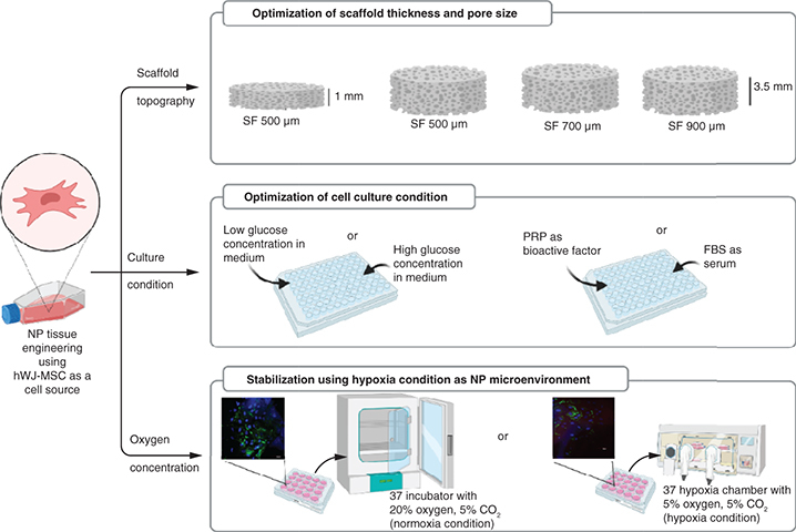
Scaffold topography and culture medium conditions for human wharton's jelly mesenchymal stem cells (hWJ-MSC) are critical components of the approach to nucleus pulposus (NP) tissue engineering. Aim: To evaluate the silk fibroin (SF) scaffold topography analysis (optimal thickness and pore diameter) and to determine culture medium conditions for the growth and differentiation of hWJ-MSC. Method: hWJ-MSCs were seeded into different thicknesses and pore size diameters and grown in different concentrations of glucose, platelet rich plasma (PRP) and oxygen. The cell-seeded scaffold was evaluated for cell attachment, growth and differentiation potency. Results & discussion: The results indicated that SF scaffold with a minimum thickness 3.5 mm and pore diameter of 500 μm with cells cultured under low glucose, 10% PRP and normoxia conditions induced the growth and differentiation of hWJ-MSCs, indicated by the accumulation of glycosaminoglycans content and the presence of type II collagen, as markers of NP-like cells.
Graphical abstract
Plain Language Summary
Until recently, the best approach to replacing a degenerating nucleus pulposus (NP) remained unclear. Tissue engineering is the most current method utilized to develop 3D cultures on scaffolds that direct cells sources into NP-like cell given optimal scaffold topography and culture conditions. Human Wharton's jelly mesenchymal stem cells (hWJ-MSC) are considered suitable multipotent stem cells for NP tissue engineering. A 3D construct of silk fibroin scaffold with a suitable thickness and pore diameter can facilitate attachment, growth and differentiation of hWJ-MSC into NP-like cells. Culture conditions with low glucose concentration on medium supplemented with PRP in normal oxygen conditions enhance the NP extracellular matrix marker, an indication that these 3D constructs and culture conditions can be developed into NP-like cells.
Supplementary data
To view the supplementary data that accompany this paper please visit the journal website at:www.tandfonline.com/doi/full/10.2217/epi-2016-0184
Author contributions
N Vanawati, performed the experiment, analysis, and wrote this paper. H Judawisastra designed experiment the idea, developing the scaffold in this experiment and contributed to the final manuscript. A Barlian, I Wibowo, designed experiment the idea, verified analytical method, supervised the research, and contributed to the final manuscript
Acknowledgments
Authors are thankful to PT Fajar Mas Murni for providing Olympus Fv1200 confocal laser-scanning microscope.
Financial & competing interests disclosure
This research was funded by Indonesia Endowment Fund for Education (Lembaga Pengelola Dana Pendidikan/LPDP) and Institut Teknologi Bandung research program. The authors have no other relevant affiliations or financial involvement with any organization or entity with a financial interest in or financial conflict with the subject matter or materials discussed in the manuscript apart from those disclosed.
No writing assistance was utilized in the production of this manuscript.
Ethical conduct of research
Human umbilical cord samples were obtained from caesarean delivery at Rumah Sakit Khusus Ibu dan Anak (RSKIA), Bandung, Indonesia. All experimental methods were approved by the Medical Health Research Ethic Committee (MHREC) Faculty of Medicine, Public Health and Nursing at Universitas Gadjah Mada – Dr Sardjito General Hospital under the number KE/KF/1482/EC/2019. Participants completed an informed consent form and sampling was carried out under the supervision of health professionals and in compliance with applicable legislation.