
Abstract
Background
The complex pathophysiology of migraine is not yet clearly understood; therefore, experimental models are essential for the investigation of the processes related to migraine headache, which include cortical spreading depression (CSD) and NO donor-induced neurovascular changes. Data on the assessment of drug efficacy in these models are often limited, which prompted us to investigate a novel combined migraine model in which an effective pharmacon could be more easily identified.
Materials and methods
In vivo electrophysiological experiments were performed to investigate the effect of nitroglycerin (NTG) on CSD induced by KCl application. In addition, sumatriptan and newly synthesized neuroactive substances (analogues of the neuromodulator kynurenic acid [KYNA]) were also tested.
Results
The basic parameters of CSDs were unchanged following NTG administration; however, propagation failure was decreased compared to the controls. Sumatriptan decreased the number of CSDs, whereas propagation failure was as minimal as in the NTG group. On the other hand, both of the KYNA analogues restored the ratio of propagation to the control level.
Discussion
The ratio of propagation appeared to be the indicator of the effect of NTG. This is the first study providing direct evidence that NTG influences CSD; furthermore, we observed different effects of sumatriptan and KYNA analogues. Sumatriptan changed the generation of CSDs, whereas the analogues acted on the propagation of the waves. Our experimental design overlaps with a large spectrum of processes present in migraine pathophysiology, and it can be a useful experimental model for drug screening.
Introduction
Migraine is one of the most common neurological disorders, affecting 17% of women and 6% of men in the US.Citation1 The environmental triggers eliciting migraine headache are diverse. The characteristic phases include aura (in a third of patients), headache, nausea, and hypersensitivity. In subgroups of patients, migraine headache is a well-managed condition; however, the therapeutic efficacy of available antimigraine drugs is in many cases limited, which necessitates unabated research efforts. The two main categories in migraine therapy include the acute and the preventive (ie, prophylactic) approaches. In acute therapy, different triptans (eg, sumatriptan) and nonsteroidal anti-inflammatory drugs are mainly used, whereas calcium-channel blockers, serotonin (ie, 5-hydroxytriptamine [5-HT]) antagonists, and β-blockers are among the drugs used for prophylaxis. Even though the research related to the processes involved in the pathomechanism of migraine has been a focus for decades, these have not yet been fully elucidated. Therefore, animal experiments are essential for further investigation of the pathophysiological processes related to migraine headache, and also for testing new substances aimed at mitigation of symptoms. The different mechanisms of migraine can be investigated from various aspects by a number of experimental models based on the sensitization or the stimulation of the trigeminal system, including the investigation of cortical spreading depression (CSD) and changes in the neurovascular system induced by NO donors.Citation2
CSD is the electrophysiological correlate of migraine aura, and represents a potential trigger for headache.Citation3 It is an intense self-propagating wave of depolarization involving neuronal and glial cells in the cerebral cortex, which is independent of functional areas and arterial territories.Citation4 CSD spreads at a velocity of 3–6 mm/min and causes massive changes in ionic homeostasis. Glutamatergic mechanisms have primary roles in the propagation of CSD. Indeed, CSD cannot be induced in cases of either P/Q-type calcium channels or N-methyl-d-aspartate (NMDA) receptors being blocked in brain slices in vitro,Citation5 or when antagonists of calcium channels or NMDA receptors are used in vivo.Citation6,Citation7 In addition, CSD upregulates several receptors in hippocampal and striatal territories, such as γ-aminobutyric acid A, different cholinergic (M1/2, α1/2), 5-HT serotonergic, and D1 dopaminergic, as well as NMDA-, α-amino-3-hydroxy-5-methyl-4-isoxazolepropionic acid-and kainate-sensitive glutamatergic receptors.Citation8,Citation9 Therefore, in addition to glutamatergic mechanisms, alterations in other neurotransmitter systems participate in CSD as well. CSD also has an effect on cerebral blood flow, being associated with an initial increase in the blood flow via arteriolar dilation (mediated predominantly by NO), which subsequently turns into long-term oligemia.Citation10,Citation11
Experimental administration of NO donors, such as nitroglycerin (NTG), is a widely used method to model head pain. NTG elicits headache in both animals and humans.Citation12,Citation13 Systemic NTG administration evokes alterations in meningeal and cortical blood flow,Citation14,Citation15 increases the level of calcitonin gene-related peptide, and induces c-Fos expression in different brain areas associated with pain perception.Citation16
With the use of these migraine models, several promising substances acting via various mechanisms can be investigated. The neuroactive metabolites of the kynurenine system provide a spectrum of possibilities to interfere with different neuropathological processes.Citation17,Citation18 Following the publication of the first studies suggesting the role of kynurenines in migraine,Citation19 a subsequent series of research proved the beneficial effects of kynurenine-related pharmacological manipulations in migraine models.Citation20,Citation21 Among these substances, kynurenic acid (KYNA) has potential neuroprotective action by interfering with glutamatergic excitotoxicity. The peripheral administration of KYNA provides a number of promising results; however, these effects are limited, because of the low penetration of KYNA through the blood–brain barrier.Citation22 Therefore, structurally modified KYNA derivates with better pharmacokinetic profiles may serve as useful tools against a large spectrum of neuropathological processes.
In this study, our aim was to investigate the effect of NTG on CSD, so as to create and validate a combined experimental migraine model, enabling the investigation of antimigraine drugs and the selection of other substances affecting the pathomechanism of migraine for further examinations. Also, we applied NTG with other molecules, including sumatriptan and novel analogues of KYNA, prior to the induction of CSD to test our combined experimental migraine model and investigate the effects of these molecules in this setting.
Materials and methods
Animals and surgery
All procedures used in this study were in accordance with the guidelines of the European Communities Council Directives (2010/63/EU) and the Hungarian Act for the Protection of Animals in Research (XXVIII.tv. 32. §). The study was approved by the National Scientific Ethical Committee on Animal Experimentation (National Competent Authority in Hungary), with the license number XX./842/2016. Male Wistar rats weighing 250–300 g were used. Animals were housed in polyethylene cages at room temperature (22°C) with a 12-hour light–dark cycle (lights on at 6 am) and free access to water and rodent laboratory chow.
All procedures were carried out under deep urethane anesthesia (1.5±0.2 g/kg body weight), as it provides very stable anesthesia in rats, lasting for several hours and being optimal for CSD recordings.Citation23,Citation24 During surgery and CSD recordings, the animals breathed spontaneously, and their body temperature was maintained at 37°C±0.5°C by the use of a self-regulating heating pad (TMP 5-b; Supertech Instruments, Budapest, Hungary) and a rectal probe. A trachea cannula was inserted, and heads of the animals were fixed in a stereotaxic frame. After exposure of the skull, three holes (2 mm in diameter) were drilled on the left side under cooling with saline with 3 mm between each hole (see ). Two holes were used for electrophysiological recordings above the sensorimotor cortex (bregma [Br] 1, 2 for E1, −2, 2 for E2, indicating distances in millimeters in the anterior–posterior and the medial–lateral axes from the Br, respectively). In the third hole (Br −5, 2), the dura and arachnoid mater were carefully removed, allowing access for the chemical induction of CSD. Before the recordings, the cortex was allowed to recover for 30 minutes.
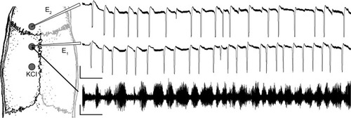
Figure 1 Experimental design and representative recording from the 2-hour period following KCl administration.
Electrophysiological recordings
Both the slow direct current (DC)-potential change and spontaneous cortical electrical activity accompanying CSD were continuously recorded for 4 hours. For the DC recordings, glass microelectrodes (1.5–2 MΩ) filled with artificial cerebrospinal fluid connected to an Ag/AgCl wire were inserted into the cortex to a depth of 800±100 μm, corresponding to cortical layer V. Electrocorticography (ECoG) was recorded from the surface of the dura with the aid of a silver electrode next to the posterior DC electrode (Br −2, 2). ECoG was recorded to monitor the depth of anesthesia and to verify the CSDs that cause a notable reduction in the ECoG amplitudes (). In both types of recordings, common reference electrodes of the same type were placed in the masticatory muscle. DC-potential changes and ECoG were recorded by connecting the electrodes to EXP-E amplifiers (Experimetria Ltd, Budapest, Hungary).
Following offline data processing with laboratory-developed software, we examined the frequency, amplitude, time to half amplitude, velocity of propagation, and propagation failure of the CSD waves. Furthermore, we compared these parameters separately between the two recording DC electrodes (E1, E2) and between the first and second hour of recording following KCl application. The velocity of CSD-wave propagation was calculated from the time required for a CSD wave to travel the distance between the two cortical recording points. This time was measured by using the beginning of the first descending phase of the slow DC-potential change as the initial point, as previously described by Batista-de-Oliveira et al.Citation25 DC shifts not exceeding the threshold of 5 mV were excluded from data analysis.
Experimental groups and treatments
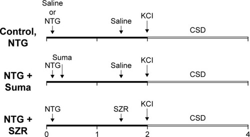
The applied pharmacons and the saline (used as control and vehicle) were injected intraperitoneally prior to KCl administration at the given time point (). The control group (n=6) received 2.5 mL of saline, whereas the NTG group (n=5) was treated with 10 mg/kg NTG (Nitro Pohl; G Pohl-Boskamp GmbH and Co KG, Hohenlockstedt, Germany) at minute 10. In the NTG + sumatriptan group (n=5), sumatriptan (Imigran; GlaxoSmithKline PLC, London, UK) at a dose of 0.6 mg/kg was applied 10 minutes after the NTG injection, in accordance with Bates et al.Citation26 The KYNA analogues (SZR-105 and SZR-109; each n=5) were both administered at a dose of 0.5 mmol/kg at minute 90 (80 minutes after NTG administration, 30 minutes before CSD induction).
Figure 2 Experimental design of the different groups. Numbers represent hours of recording.
The KYNA analogues SZR-105 and SZR-109 are novel neuroactive KYNA derivatives, substituted at C3. According to preliminary studies, these molecules might be effective neuroprotective or effective compounds in certain neurological diseases (eg, in Alzheimer’s disease, Parkinson’s disease, Huntington’s disease, amyotrophic lateral sclerosis, stroke, multiple sclerosis, and migraine).
In all groups, CSD waves were continuously elicited for 2 hours from the 120th min by placing a cotton ball (diameter 1 mm) soaked with 1 M KCl solution onto the cortex through the posterior hole. To keep the cotton ball moist, it was impregnated with 5 μL of KCl solution every 15 minutes.
Statistical analysis
IBM SPSS 20 software was used for the analyses. Electro-physiological data between the two electrodes were compared separately by the use of nonparametric related-sample Friedman two-way analysis of variance (ANOVA) by rank. To analyze the differences between the groups, nonparametric independent-sample Kruskal–Wallis ANOVA was used. Data are expressed as mean and standard deviation. Differences were considered statistically significant when P≤0.05.
Results
The slow potential change in the DC-potential recordings clearly denoted the presence of CSDs after KCl stimulation, which could also be easily detected by the farther, second DC electrode (E2). In some cases, more CSDs were detected by E2, which can be attributed to a very slight depolarization of the area under E1.
After analysis of the data, the amplitude, velocity, and time to half amplitude of the CSDs showed no significant differences between the groups (). None of these parameters differed significantly when comparing the data obtained from the two electrodes separately () or when comparing the first and the second hour (data not shown). However, a slight increase in the amplitudes was observed at E2.
Table 1 Parameters of CSD
In the control group, we observed a constantly occurring propagation failure (20%±1%) (), which resulted in decreased CSD numbers recorded by E2. This tendency was observed in almost all the recordings (), and proved to be significant (P=0.046) based on pairwise comparison (two-way Friedman’s ANOVA). The CSD numbers (similarly to all other groups) recorded by either E1 or E2 showed no difference between the first and second hours of KCl administration (E1 9.3±4, 10.5±4; E2 8.0±3, 7.4±2) ().
Figure 3 CSD frequency and ratio of propagation.
Abbreviations: CSD, cortical spreading depression; NTG, nitroglycerin; ANOVA, analysis of variance; SD, standard deviation; Suma, sumatriptan.
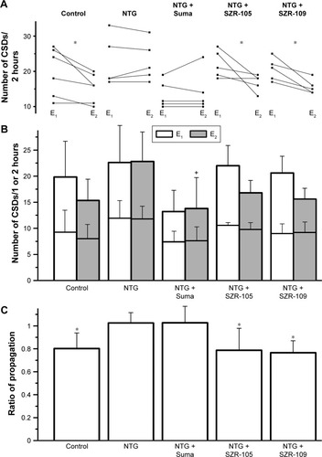
The administration of NTG slightly but insignificantly elevated CSD numbers and significantly changed the ratio of propagation (1.02±0.1). In this case, no significant difference was observed between the electrodes, as opposed to the control group (). In the NTG + sumatriptan group, CSD numbers decreased when sumatriptan was applied 10 minutes after NTG treatment. Significant differences were detectable in multiple comparisons of CSD numbers recorded by E2 in the different groups (). In this group, the ratio of propagation (1.03±0.1) was similar to that in the NTG group ().
Both of the KYNA analogues applied after NTG injection restored CSD numbers and the ratio of propagation to the control level (). In each animal (n=5 in both KYNA analogue-treated groups), the number of CSDs at E2 was less than or at most equal with that observed at E1, which resulted in significant decreases in related-sample analyses (P=0.046 and P=0.025 for the two KYNA-analogue groups, respectively; ).
Discussion
Despite the large amount of accumulated knowledge about the processes involved in migraine pathogenesis, the available treatments are at present often limited and suboptimal. Therefore, new experimental models, candidate compounds and drug targets are still the focus of antimigraine research.Citation27,Citation28 Cortical excitability in CSD is a physiological correlate of aura in migraine,Citation29,Citation30 which prompted us to investigate the hypothesis whether NTG can influence the characteristics of CSDs.
Based on a number of studies, CSD is an “all or none” phenomenon, making it difficult to detect remarkable changes in various CSD parameters following the acute administration of potential antimigraine drugs.Citation31–Citation33 The most relevant CSD parameters include the susceptibility (examined by increasing electrical stimulation), the frequency (in cases of continuous KCl application), and the velocity of SD. Of note, the amplitude and the duration are quite constant in most experiments.Citation34
In the present study, the progression of the depolarization wave failed between the two recording points in 20% of the CSDs, indicative of the presence of inhibitory effects in the cortical tissue under physiological conditions (notably during anesthesia). However, the propagation of the CSDs was unimpeded after the systemic administration of NTG, molecules of which may provide support against homeostatic and energetic disturbances caused by the CSDs,Citation35 resulting in enhanced propagation. In contrast to our results, some studies have proved an inverse relationship between tissue NO level and susceptibility in human and rodent corticesCitation36 and also in chicken retina.Citation37 Low NO level or inhibited NOS increased tissue susceptibility to CSD. Furthermore, CSDs themselves increase NO levels,Citation38 and thus additional NTG may contribute to the release or appearance of other NO metabolites. Further investigations are needed to identify the detailed mechanisms and to explore the active substances that take part in this phenomenon. Nevertheless, in point of fact, we suggest that the altered physiological state caused by NTG administration can be influenced more easily than the baseline parameters of CSDs; therefore, this model may provide an opportunity to test compounds with only moderate or subtle effects via the assessment of the ratio of propagation. Our results clearly demonstrate the effect of NTG administration on CSD propagation. There have been limited studies to investigate the NTG effect on CSD. Read et al confirmed that NTG causes elevated brain NO levels in anesthetized cats.Citation39 The susceptibility, duration, and amplitude of CSDs were not changed by NTG; however, CSD-induced local NO concentration was elevated. In another study, Bates et al performed experiments on mice to investigate CSD susceptibility following NTG administration, but they did not find any differences between the groups.Citation26 In our study, sumatriptan decreased the frequency of CSDs; however, the ratio of propagation remained similar to that seen in the NTG group. Sumatriptan has been proven to be effective in migraine by acting on the serotonin system through the activation of 5-HT1B/1D receptors. Decreased 5-HT level is closely associated with headache.Citation40 It is known that 5-HT receptors are involved in neurogenic cranial vascular changes. Indeed, meningeal blood flow increases following the application of the 5-HT7-receptor agonist, whereas 5-HT7-receptor antagonist plus sumatriptan as a 5-HT1B/1D agonist causes decreased blood flow.Citation41 In a prior study, sumatriptan decreased the concentration of NO following CSD induction,Citation42 and alleviated mechanical and thermal allodynia that developed as a result of NTG administration.Citation26 In a vessel-free preparation of chicken retina, sumatriptan blocked the SD in a concentration-dependent manner, which indicates that the effect must be related to neuronal tissue.Citation43 Nevertheless, there is no evidence that sumatriptan can strongly modulate CSDs.Citation44,Citation45 In the present study, we cannot give a full explanation of the mechanisms underlying our results. However, we suggest that the 5-HT receptors directly involved in CSD could contribute to this effect.
This is the first study with these novel KYNA analogues demonstrating an effect; indeed, the elevated ratio of propagation was restored to the control level by these compounds, whereas the number of CSDs remained largely unchanged. There are several possibilities to explain these results, including the potential modulatory effect of these analogues on glutamatergic transmission by means of the inhibition of NMDA receptors, similarly to KYNA; however, further investigations are needed to be conclusive at this point. The beneficial effects of KYNACitation46 and its different analogues have been proven in different migraine models. The KYNA analogue SZR-72 mitigated systemic NTG-induced increase in the number of neurons expressing c-FosCitation47 and nNOS.Citation48 Furthermore, SZR-72 attenuated the NTG-induced decrease of histochemical changes in the caudal trigeminal nucleus.Citation49 The dissimilar effects of sumatriptan and KYNA analogues on CSDs suggest two inhibitory mechanisms. Further investigations are required to clarify the mechanisms underlying these effects; however, we can conclude that sumatriptan acts on the generation of the waves, whereas KYNA analogues interfere with the propagation of CSDs. Nevertheless, we cannot exclude the possibility that the analogues may counteract the effect of NTG and thereby restore normal conditions.
Conclusion
In summary, peripheral administration of NTG enhanced the propagation of CSDs. Sumatriptan, a widely used antimigraine drug, caused detectable changes in CSD frequency, which indicates the relevance of our model. Additionally, the elevated ratio of CSD propagation caused by NTG was decreased by SZR-105 and SZR-109. These results clearly show the effectiveness of these promising KYNA analogues. Here, we have presented a combined experimental migraine model for testing drugs and other pharmacological agents affecting processes involved in the pathomechanism of migraine.
Acknowledgments
This work was supported by projects OTKA KI05077 and TÁMOP-4.2.2.A-11/1/KONV-2012-0052, the European Union and the state of Hungary, cofinanced by the European Social Fund in the framework of TÁMOP 4.2.4.A/2-11-1-2012-0001 National Excellence Program, by the MTA-SZTE Neuroscience Research Group, by the Hungarian Brain Research Program (grant KTIA_13_NAP-A-III/9), by GINOP-2.3.2-15-2016-00034 and by the EUROHEADPAIN (FP7-Health 2013 Innovation, grant 602633). The authors gratefully thank Dr Levente Szalárdy for linguistic correction of the manuscript.
Disclosure
The authors report no conflicts of interest in this work.
References
- LiptonRBBigalMEToward an epidemiology of refractory migraine: current knowledge and issues for future researchHeadache2008486 791 79818479422
- Jansen-OlesenITfelt-HansenPOlesenJAnimal migraine models for drug development: status and future perspectivesCNS Drugs20132712 1049 106824234657
- AyataCCortical spreading depression triggers migraine attack: proHeadache2010504 725 73020456160
- SomjenGGAristides Leão’s discovery of cortical spreading depressionJ Neurophysiol2005941 2 415985690
- TotteneAUrbaniAPietrobonDRole of different voltage-gated Ca2+ channels in cortical spreading depression: specific requirement of P/Q-type Ca2+ channelsChannels (Austin)201152 110 11421150292
- PeetersMGunthorpeMJStrijbosPJGoldsmithPUptonNJamesMFEffects of pan- and subtype-selective N-methyl-D-aspartate receptor antagonists on cortical spreading depression in the rat: therapeutic potential for migraineJ Pharmacol Exp Ther20073212 564 57217267580
- RichterFEbersbergerASchaibleHGBlockade of voltage-gated calcium channels in rat inhibits repetitive cortical spreading depressionNeurosci Lett20023342 123 12612435487
- AyataCSpreading depression: from serendipity to targeted therapy in migraine prophylaxisCephalalgia20092910 1095 111419735536
- HaghirHKovacSSpeckmannEJZillesKGorjiAPatterns of neurotransmitter receptor distributions following cortical spreading depressionNeuroscience20091634 1340 135219665048
- ColonnaDMMengWDealDDBusijaDWNitric oxide promotes arteriolar dilation during cortical spreading depression in rabbitsStroke19942512 2463 24707526490
- PiperRDLambertGADuckworthJWCortical blood flow changes during spreading depression in catsAm J Physiol19912611 Pt 2 H96 H1021858935
- OlesenJThomsenLLIversenHNitric oxide is a key molecule in migraine and other vascular headachesTrends Pharmacol Sci1994155 149 1537538702
- TassorelliCJosephSASystemic nitroglycerin induces Fos immunoreactivity in brainstem and forebrain structures of the ratBrain Res19956821–2 167 1817552308
- GrecoRMeazzaCMangioneASTemporal profile of vascular changes induced by systemic nitroglycerin in the meningeal and cortical districtsCephalalgia2011312 190 19820693231
- StreckerTDuxMMesslingerKNitric oxide releases calcitonin-gene-related peptide from rat dura mater encephali promoting increases in meningeal blood flowJ Vasc Res2002396 489 49612566974
- TassorelliCJosephSANADPH-diaphorase activity and Fos expression in brain nuclei following nitroglycerin administrationBrain Res19956951 37 448574645
- SchwarczRBrunoJPMuchowskiPJWuHQKynurenines in the mammalian brain: when physiology meets pathologyNat Rev Neurosci2012137 465 47722678511
- VecseiLSzalardyLFulopFToldiJKynurenines in the CNS: recent advances and new questionsNat Rev Drug Discov2013121 64 8223237916
- Knyihár-CsillikEChadaideZOkunoEKynurenine aminotransferase in the supratentorial dura mater of the rat: effect of stimulation of the trigeminal ganglionExp Neurol20041862 242 24715026260
- Knyihár-CsillikEToldiJKrisztin-PévaBPrevention of electrical stimulation-induced increase of c-Fos immunoreaction in the caudal trigeminal nucleus by kynurenine combined with probenecidNeurosci Lett20074182 122 12617382471
- Knyihár-CsillikEToldiJMihályAKynurenine in combination with probenecid mitigates the stimulation-induced increase of c-Fos immunoreactivity of the rat caudal trigeminal nucleus in an experimental migraine modelJ Neural Transm (Vienna)20071144 417 42116897600
- SalvatiPUkmarGDhoLBrain concentrations of kynurenic acid after a systemic neuroprotective dose in the gerbil model of global ischemiaProg Neuropsychopharmacol Biol Psychiatry1999234 741 75210390731
- GorelovaNAKrivánekJBuresJFunctional and metabolic correlates of long series of cortical spreading depression waves in ratsBrain Res19874041–2 379 3813032340
- KudoCNozariAMoskowitzMAAyataCThe impact of anesthetics and hyperoxia on cortical spreading depressionExp Neurol20082121 201 20618501348
- Batista-de-OliveiraMLopesAAMendes-da-SilvaRFGuedesRCAging-dependent brain electrophysiological effects in rats after distinct lactation conditions, and treadmill exercise: a spreading depression analysisExp Gerontol2012476 452 45722504277
- BatesEANikaiTBrennanKCSumatriptan alleviates nitroglycerin-induced mechanical and thermal allodynia in miceCephalalgia2010302 170 17819489890
- GuptaSVillalónCMThe relevance of preclinical research models for the development of antimigraine drugs: focus on 5-HT(1B/1D) and CGRP receptorsPharmacol Ther20101281 170 19020655327
- LinkASKurisAEdvinssonLTreatment of migraine attacks based on the interaction with the trigemino-cerebrovascular systemJ Headache Pain200891 5 1218217201
- BowyerSMAuroraKSMoranJETepleyNWelchKMMagnetoencephalographic fields from patients with spontaneous and induced migraine auraAnn Neurol2001505 582 58711706963
- HadjikhaniNDel RioMSWuOMechanisms of migraine aura revealed by functional MRI in human visual cortexProc Natl Acad Sci U S A2001988 4687 469211287655
- AyataCJinHKudoCDalkaraTMoskowitzMASuppression of cortical spreading depression in migraine prophylaxisAnn Neurol2006594 652 66116450381
- CostaCTozziARaineroICortical spreading depression as a target for anti-migraine agentsJ Headache Pain201314 6223879550
- Eikermann-HaerterKLeeJHYalcinNMigraine prophylaxis, ischemic depolarizations, and stroke outcomes in miceStroke2015461 229 23625424478
- Eikermann-HaerterKCanAAyataCPharmacological targeting of spreading depression in migraineExpert Rev Neurother2012123 297 30622364328
- WangMObrenovitchTPUrenjakJEffects of the nitric oxide donor, DEA/NO on cortical spreading depressionNeuropharmacology2003447 949 95712726826
- PetzoldGCHaackSvon Bohlen und HalbachONitric oxide modulates spreading depolarization threshold in the human and rodent cortexStroke2008394 1292 129918309156
- UlmerHJde LimaVMHankeWEffects of nitric oxide on the retinal spreading depressionBrain Res19956911–2 239 2428590060
- ReadSJSmithMIHunterAJUptonNParsonsAASB-220453, a potential novel antimigraine agent, inhibits nitric oxide release following induction of cortical spreading depression in the anaesthetized catCephalalgia2000202 92 9910961764
- ReadSJSmithMIHunterAJParsonsAAEnhanced nitric oxide release during cortical spreading depression following infusion of glyceryl trinitrate in the anaesthetized catCephalalgia1997173 159 1659170338
- LambertGAHoskinKLZagamiASCortico-NRM influences on trigeminal neuronal sensationCephalalgia2008286 640 65218454788
- WangXFangYLiangJYanMHuRPanX5-HT7 receptors are involved in neurogenic dural vasodilatation in an experimental model of migraineJ Mol Neurosci2014542 164 17024584680
- ReadSJParsonsAASumatriptan modifies cortical free radical release during cortical spreading depression: a novel antimigraine action for sumatriptan?Brain Res20008701–2 44 5310869500
- Maranhão-FilhoPAMartins-FerreiraHVincentMBRibeiroLJNovisSASumatriptan blocks spreading depression in isolated chick retinaCephalalgia1997178 822 8259453269
- IngvardsenBKLaursenHOlsenUBHansenAJPossible mechanism of c-Fos expression in trigeminal nucleus caudalis following cortical spreading depressionPain1997723 407 4159313281
- BradleyDPSmithMINetsiriCDiffusion-weighted MRI used to detect in vivo modulation of cortical spreading depression: comparison of sumatriptan and tonabersatExp Neurol20011722 342 35311716558
- OláhGHerédiJMenyhártAUnexpected effects of peripherally administered kynurenic acid on cortical spreading depression and related blood-brain barrier permeabilityDrug Des Devel Ther2013167 981 987
- Knyihar-CsillikEMihalyAKrisztin-PevaBThe kynurenate analog SZR-72 prevents the nitroglycerol-induced increase of c-Fos immunoreactivity in the rat caudal trigeminal nucleus: comparative studies of the effects of SZR-72 and kynurenic acidNeurosci Res2008614 429 43218541319
- VámosEPárdutzAVargaHL-Kynurenine combined with probenecid and the novel synthetic kynurenic acid derivative attenuate nitroglycerin-induced nNOS in the rat caudal trigeminal nucleusNeuropharmacology2009574 425 42919580819
- VámosEFejesAKochJKynurenate derivative attenuates the nitroglycerin-induced CamKIIα and CGRP expression changesHeadache2010505 834 84319925620