
Abstract
A gene drug delivery system for glioma therapy based on transferrin (Tf)-modified polyamidoamine dendrimer (PAMAM) was prepared. Gene drug, tumor necrosis factor-related apoptosis-inducing ligand (hTRAIL)-encoding plasmid open reading frame (pORF-hTRAIL, Trail), was condensed by Tf-modified PAMAM to form nanoparticles (NPs). PAMAM-PEG-Tf/DNA NPs showed higher cellular uptake, in vitro gene expression, and cytotoxicity than PAMAM-PEG/DNA NPs in C6 cells. The in vivo targeting efficacy of NPs was visualized by ex vivo fluorescence imaging. Tf-modified NPs showed obvious glioma-targeting trend. Plasmid encoding green fluorescence protein (GFP) was also condensed by modified or unmodified PAMAM to evaluate the in vivo gene expression level. The PAMAM-PEG-Tf/plasmid encoding enhanced green fluorescence protein (pEGFP) NPs exhibited higher GFP expression level than PAMAM-PEG/pEGFP NPs. TUNEL assay revealed that Tf-modified NPs could induce much more tumor apoptosis. The median survival time of PAMAM-PEG-Tf/Trail-treated rats (28.5 days) was longer than that of rats treated with PAMAM-PEG/Trail (25.5 days), temozolomide (24.5 days), PAMAM-PEG-Tf/pEGFP (19 days), or saline (17 days). The therapeutic effect was further confirmed by magnetic resonance imaging. This study demonstrated that targeting gene delivery system had potential application for the treatment of glioma.
Keywords:
Introduction
Malignant gliomas, which have higher morbidity among brain tumors of adults, have a median survival time of approximately 12–15 months.Citation1 The prognosis of this brain tumor remains poor due to the insufficient response to the available treatments, including surgical intervention, radiation, and chemotherapy.Citation2 One of the obstacles for glioma therapies is the infiltrative nature of glioma cells, which could invade into normal brain parenchyma. Due to the importance of normal brain function, excessive surgical resection is restricted which could lead to residual of tumor cells.Citation3,Citation4 Another obstacle is the non-specificity of adjuvant treatment modalities including radiotherapy and chemotherapy, which cannot specifically reach the widely spread tumor cells. These modalities are also related to toxicities in terms of both systemic illness and cognitive impairment.Citation5 Thus, new approaches are critical to be developed to treat this devastating disease with reduced side effects.
The tumor necrosis factor-related apoptosis-inducing ligand (TRAIL) could induce apoptosis via the p53-independent pathway by binding to the death receptors 4 (TRAIL-R1) and 5 (TRAIL-R2/KILLER) at the cell surface.Citation6 The specificity of cytotoxicity could be achieved by the differences of receptor expressions. On one hand, many glioma cells express the agonist TRAIL receptors with no or undetectable levels of the antagonist receptors.Citation7 On the other hand, normal cells also express antagonist TRAIL receptors.Citation8,Citation9 Therefore, TRAIL protein may allow killing tumor cells alone without affecting the normal brain cells. TRAIL protein has been reported to induce tumor regression in a glioma xenograft model via intravenous administration.Citation10,Citation11 However, some disadvantages, including the short half-life, immunogenicity, and enzymolysis, limit the usage of protein drugs. Gene therapy with genes encoding therapeutic proteins like TRAIL may offer therapeutic promise including long-lasting expression of therapeutic proteins.
Due to the invasive nature, tumor cells could penetrate into normal brain tissue. The tumor vasculatures in the infiltrated region possess some features of normal brain including blood–brain barrier (BBB).Citation12–Citation14 Gene drugs cannot reach these tumor regions due to the impermeability of BBB to the large molecules.Citation15–Citation18 Therefore, there is an urgent need for developing smart carriers for gene delivery targeting glioma. Learning from the nature, many specific receptors including transferrin (Tf) receptor and insulin receptor are overexpressed on BBB that can potentially be utilized for facilitating brain targeting.Citation19–Citation21 Among these receptors, Tf receptor is a very promising one because of the restricted expression in brain capillaries than other tissues.Citation22 Besides, the expression level of Tf receptor on tumor cells is much higher than that of normal cells.Citation23,Citation24 Thus, Tf is selected as the glioma-targeting ligand.
Non-viral vectors have been used to deliver therapeutic genes into the brain with higher safety. Among the vectors reported, polyamidoamine dendrimer (PAMAM) has proven to be a promising gene carrier.Citation25 PAMAM-PEG-Tf has been successfully synthesized previously and showed great brain-targeting efficiency.Citation26 Besides, it has been reported that Tf-conjugated polymersomes achieved preferential C6 glioma targeting in vivo and Tf-modified cisplatin liposome were identified to accumulate in malignant glioma cells in bEnd3/C6 co-culture BBB models.Citation27,Citation28
In this study, hTRAIL-encoding plasmid (pORF-hTRAIL, Trail) was selected as the therapeutic gene for glioma treatment. PAMAM-PEG-Tf was synthesized, and incorporated with pORF-hTRAIL to form nanoparticles (NPs). The antitumor effect of the NPs was evaluated in C6 glioma tumors-bearing Sprague Dawley (SD) rats.
Materials and methods
The plasmids encoding enhanced green fluorescence protein N2 (pEGFP-N2; Clontech, Palo Alto, CA, USA) and pORF-hTRAIL (Invitrogen, San Diego, CA, USA) were purified by QIAGEN Plasmid Mega Kit (Qiagen GmbH, Hilden, Germany). Human holo-Tf and methyl green were purchased from Sigma-Aldrich (St Louis, MO, USA). PAMAM G5 dendrimer was purchased from Dendritech, Inc (Midland, MI, USA). A-Maleimidyl-u-N-hydroxysuccinimidyl polyethyleneglycol (NHS-PEG-MAL, molecular weight 3,500) was obtained from Jenkem Technology (Beijing, People’s Republic of China). 4,6-diamidino-2-phenylindole (DAPI) and ethidium monoazide bromide (EMA) were purchased from Molecular Probes (Eugene, OR, USA).
Male SD rats (4–5 weeks) of 200–220 g body weight were purchased from Sino-British SIPPR/BK Lab Animal Ltd. (Shanghai, People’s Republic of China) and maintained under standard housing conditions. All animal experimental protocols were in accordance with guidelines evaluated and approved by the ethics committee of Zhejiang University.
The rat C6 glioma cell line was kindly provided by Prof Feng L (Institute of Neuroscience, Shanghai Institutes for Biological Sciences). In addition, this cell line was purchased from Shanghai Cell Bank, Chinese Academy of Medical Sciences, Beijing, People’s Republic of China. C6 cells were expanded and maintained in Dulbecco’s Modified Eagle’s Medium (DMEM) supplemented with 10% heat-inactivated fetal calf serum, 100 U/mL penicillin, and 100 mg/mL streptomycin and cultured at 37°C under a humidified atmosphere containing 5% CO2.
Synthesis of PAMAM derivatives
Synthesis of PAMAM derivate was carried out as previously reported.Citation26 In brief, PAMAM was reacted with NHS-PEG-MAL at a mole ratio of 1:2 in phosphate-buffered saline (PBS) pH 8.0 for 2 hours at room temperature (RT). The primary amino groups of PAMAM were specifically reacted with NHS groups of NHS-PEG-MAL. The resulting conjugate, PAMAM-PEG-MAL, was purified by ultrafiltration (cutoff: 5 kDa) and the solution was changed to PBS 7.0. Tf was thiolated by the Traut’s reagent, and the extent of thiolation was analyzed by the Ellmann’s reagent. Then PAMAM-PEG-MAL was reacted with Tf at a mole ratio of 1:1 in PBS 7.0 for 24 hours at RT. The MAL groups of PAMAM-PEG-MAL were specifically reacted with thiol groups of Tf. Theoretically, there are totally 128 free amine groups in G5 PAMAM and two of them were modified with polyethyleneglycol (PEG). One molecular Tf was attached to the end of one PEG.
Preparation and characterization of dendrimers/DNA NPs
Dendrimers (PAMAM-PEG-Tf) were diluted to desired concentrations in PBS 7.4. DNA solution was added to obtain appropriate weight ratios (0.1:1, 0.5:1, 1:1, 3:1, PAMAM to pORF-hTRAIL) and then vortexed for 30 seconds at RT. Agarose gel electrophoresis was performed to evaluate the retardation of DNA in the NPs. Freshly prepared NPs with weight ratio of 3:1 were used in the subsequent experiments. The morphology of NPs was examined under a transmission electron microscope (TEM) (JEM-2010, JEOL, Tokyo, Japan).
Cellular uptake of NPs in C6 cells
Plasmid DNA (pORF-hTRAIL) was covalently labeled with a fluorescent dye, EMA. Fresh plasmid DNA solution (0.1 mg/mL) and EMA (1 mg/mL) were incubated at equal volume for 0.5 hour in the dark. The complex was then exposed to ultraviolet (UV) light (365 nm) for 1 hour. The resulting solution was precipitated by adding ethanol to a final concentration of 30% (v/v). The precipitate was collected and dissolved in 50 mM sodium sulfate solution. This EMA-DNA solution was used to prepare the NPs as described earlier.
C6 cells were seeded at a density of 5×10³/well in 24-well plates (Corning-Coaster, Tokyo, Japan), incubated for 48 hours, and checked under the microscope for morphology. When reaching 90% confluence, cells were incubated with naked DNA (EMA-labeled pORF-hTRAIL, Trail), PAMAM-PEG/Trail, and PAMAM-PEG-Tf/Trail at a dose of 5 µg/well DNA. After 2-hour incubation, the medium was removed, and the cells were washed twice with PBS. Then, the cells were visualized and imaged using a fluorescence microscope.
In vitro gene expression
C6 cells were seeded at a density of 5×10³/well in 24-well plates, incubated for 24 hours, and checked under the microscope for morphology. When reaching 60% confluence, cells were incubated with PBS, naked DNA (pEGFP-N2), PAMAM-PEG/pEGFP, and PAMAM-PEG-Tf/pEGFP at a dose of 5 µg/well DNA. After 2-hour incubation, the medium was removed, and the cells were washed twice with PBS. After incubation for another 48 hours, cells were washed and imaged using a fluorescence microscope.
In vitro cell apoptosis
C6 cells were seeded at a density of 1×10⁴/well in six-well plates, incubated for 24 hours, and checked under the microscope for morphology. When reaching 60% confluence, cells were incubated with PBS, naked DNA (Trail), PAMAM-PEG/Trail, and PAMAM-PEG-Tf/Trail at a dose of 10 µg/well DNA. After 2-hour incubation, cells were washed and further cultured in DMEM medium containing 10% fetal bovine serum for 48 hours. Then, the cells were incubated with Annexin V-fluorescein isothiocyanate (FITC) Apoptosis Detection Kit following the manufacturer’s protocol (KeyGEN, Nanjing, People’s Republic of China). Briefly, the collected cells were washed twice with PBS and incubated with Annexin V-FITC and propidium iodide for 15 minutes in the dark at RT. The cells were washed twice and then analyzed within 60 minutes by flow cytometry (BD FACSCalibur™; BD Biosciences, San Jose, CA, USA).
In vivo distribution of NPs in C6 glioma xenografted SD rat brain
C6 model: C6 cells (1×10⁵ per well) were cultured in petri dish until 90% confluence was reached. Cells (2×10⁴/5 µL in PBS) were implanted into the right striatum (3 mm lateral, 1 mm before the bregma, and 5 mm of depth) of SD rats by using a stereotactic fixation device with a rat adaptor. Eight days after implantation, rats received PAMAM-PEG/Trail and PAMAM-PEG-Tf/Trail NPs via the tail vein at a dose of 200 µg pDNA/rat, and 2 hours later were anesthetized with diethyl ether and killed by decapitation. Distribution of NPs in brain was investigated using Maestro™ (PerkinElmer Inc., Waltham, MA, USA) 2 in vivo imaging system 2 hours after injection.
Qualitative evaluation of in vivo gene expression
The PAMAM-PEG/pEGFP and PAMAM-PEG-Tf/pEGFP NPs (10:1, PAMAM to DNA, w/w) were injected into the tail vein of rats at a dose of 200 mg DNA/rat 10 days after C6 implantation. After 48 hours, animals were anesthetized with 10% chloral hydrate and sacrificed by the perfusion of saline and 4% paraformaldehyde. The brains were fixed in 4% paraformaldehyde and dehydrated in 15% and 30% sucrose solution. Frozen sections of rat gliomas with 20 µm thickness were prepared with a cryotome Cryostat (Leica, CM 1900, Leica Microsystems, Wetzlar, Germany) and stained with 300 nM DAPI for 15 minutes at RT. After washing twice with PBS, the sections were examined under the fluorescence microscope.
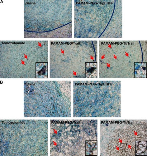
Immunohistochemistry of apoptosis markers
At day 8, 10, 12, 14, and 16 after implantation, rats received PAMAM-PEG/Trail, PAMAM-PEG-Tf/pEGFP, PAMAM-PEG-Tf/Trail NPs, or saline (control) via intravenous at a dose of 200 µg DNA/rat. Temozolomide was administrated via intragastric administration at day 8, 9, 10, 11, and 12 (one course of treatment). At day 18 after implantation, rats were sacrificed and used to prepare brain glioma frozen sections (20 µm) as described earlier. Terminal deoxynucleotidyl transferase-Biotin-dUTP nick end labeling (TUNEL) was used to detect apoptosis.
Therapeutic effect evaluation by survival monitoring and MRI imaging
After the same treatment course as described earlier, the survival of rats was monitored every day (n=10). Magnetic resonance imaging (MRI) was performed using a Bruker Biospec 4.7 T/30 cm scanner as previously suggested.Citation29 Imaging was performed using a custom-built 50 mm diameter send-receive birdcage volume coil. Gradients were 12 cm diameter at 25 G/cm. Heartbeat rates were monitored so as to keep it at 60–120 times per minute by adjusting the ratio of isoflurane/oxygen. T2-weighted images were performed with the following parameters: slice thickness =1.00 mm, FOV (field of view) =2 cm ×2 cm, echo time =11 ms, repetition time =3,000 ms, number of averages =8, and matrix size =128×128. MRI was acquired before and after the same therapeutic regimen mentioned earlier.
Results
Preparation of dendrimers/DNA NPs
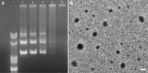
The ability of PAMAM-PEG-Tf to condense DNA was evaluated by electrophoretic analysis at different weight ratios. Complete retardation was achieved at the weight ratio of 3:1 (). This formulation was used in the following studies. The TEM results showed a narrow size distribution of NPs and the average diameter was 119±13 nm, data is presented as mean ± standard deviation ().
Figure 1 Characterization of dendrimers/DNA NPs.
Abbreviations: NP, nanoparticle; PAMAM, polyamidoamine dendrimer; PEG, polyethyleneglycol; Tf, transferrin.
Cellular uptakes of NPs
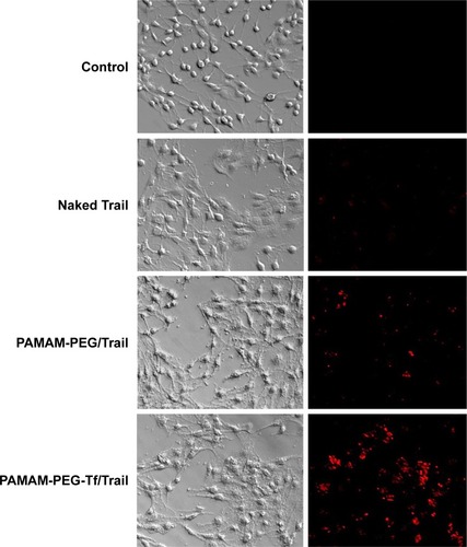
Trail was previously labeled with EMA for intracellular tracking. The cellular uptake of naked Trail, PAMAM-PEG/Trail, and PAMAM-PEG-Tf/Trail into C6 cells was compared by fluorescent imaging (). Because of the negative charge, naked Trail showed little uptake into cells. When condensed into NPs, the cellular uptake was greatly increased. Besides, targeting NPs PAMAM-PEG-Tf/Trail exhibited significantly higher uptake than non-targeting NPs PAMAM-PEG/Trail. Tf conjugation facilitated the cellular uptake of NPs in a specific manner.
Figure 2 Cellular uptake of NPs in C6 cells.
Abbreviations: EMA, ethidium monoazide bromide; NP, nanoparticle; PAMAM, polyamidoamine dendrimer; PEG, polyethyleneglycol; Tf, transferrin.
In vitro gene expression
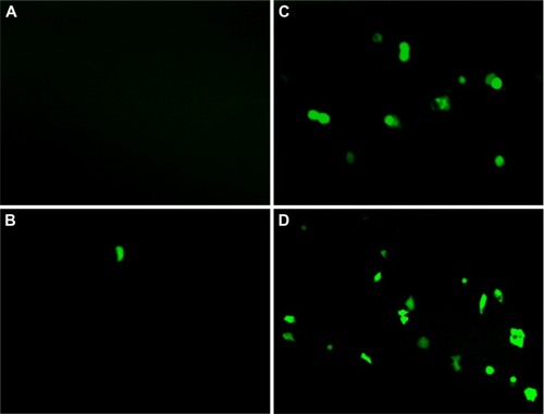
The expression of pEGFP was utilized to demonstrate the transfection efficiency of NPs. Due to the poor cellular uptake, naked pEGFP-treated cells showed little green fluorescence protein (GFP) expression as the blank control (). NP-treated cells had more GFP expression than naked pEGFP-treated ones, which contributed to the condensation of positively charged PAMAM (). Tf modification further increased GFP expression due to the overexpression of Tf receptors, which resulted in enhanced cellular uptake.
Figure 3 In vitro gene expression of NPs in C6 cells.
Abbreviations: NP, nanoparticle; PBS, phosphate-buffered saline; PAMAM, polyamidoamine dendrimer; PEG, polyethyleneglycol; pEGFP, plasmid encoding enhanced green fluorescence protein; Tf, transferrin.
In vitro cell apoptosis
When the transfection efficiency was confirmed, the cell apoptosis of NPs containing therapeutic gene was evaluated by Annexin V-FITC staining. Annexin V-FITC could label the membrane phospholipid phosphatidylserine, which is translocated from the inner to the outer side of cell membranes in early cell apoptosis. Propidium iodide is a molecular probe that can penetrate through destructed membranes and label nucleus.Citation30 UL stands for dead cells, UR for late apoptosis cells, LL for live cells, and LR for early apoptotic cells. Naked Trail induced total apoptosis rate (UR + LR) similar to that of PBS-treated group (). PAMAM-PEG/Trail-treated group had a total apoptosis rate of 19.22% (). In addition, PAMAM-PEG-Tf/Trail induced most significant cell apoptosis of 40.24% (), which demonstrated the superiority of dendrimer condensing and Tf modification.
Figure 4 In vitro cell apoptosis.
Abbreviations: PBS, phosphate-buffered saline; PAMAM, polyamidoamine dendrimer; PEG, polyethyleneglycol; Tf, transferrin; FITC, fluorescein isothiocyanate; UL, upper left; UR, upper right; LL, lower left; LR, lower right.

In vivo distribution of NPs in C6 glioma-bearing rat brain
C6 glioma-bearing rats were injected with PAMAM-PEG-Tf/EMA-labeled DNA NPs and PAMAM-PEG/DNA NPs. Ex vivo fluorescent images of brain were taken 2 hours after injection. EMA-labelled DNA was obviously accumulated in the brain tumor, which treated with PAMAM-PEG-Tf/DNA NPs (), while that in the brain tumor treated with PAMAM/DNA NPs was less concentrated ().
Figure 5 In vivo distribution of NPs in C6 glioma model.
Abbreviations: NP, nanoparticle; PAMAM, polyamidoamine dendrimer; PEG, polyethyleneglycol; Tf, transferrin.
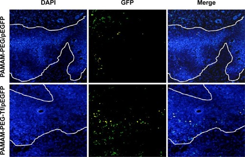
Qualitative evaluation of in vivo gene expression
GFP expression in the brain tumor was examined 48 hours after administration (). In PAMAM-PEG/pEGFP NP-treated group, GFP was expressed near the margin of the glioma region. In PAMAM-PEG-Tf/pEGFP NP-treated group, GFP was generally found both near the margin and inside of the tumor and was much more than that of PAMAM-PEG/pEGFP NP-treated group.
Figure 6 Qualitative evaluation of in vivo gene expression.
Abbreviations: DAPI, 4,6-diamidino-2-phenylindole; PAMAM, polyamidoamine dendrimer; pEGFP, plasmid encoding enhanced green fluorescence protein; PEG, polyethyleneglycol; Tf, transferrin.
Immunohistochemistry of apoptosis markers
DNA fragmentation, a marker of late apoptosis, in nuclei of tumor cells was detected in margin region () and central region () using TUNEL assay. Virtually, little apoptotic cells were detected after PAMAM-PEG-Tf/pEGFP NPs and saline treatments in neither margin nor central region. PAMAM-PEG/Trail NPs and Temozolomide showed comparable apoptosis levels in margin region, but little tumor apoptosis was found in the central region. PAMAM-PEG-Tf/Trail NPs induced most tumor apoptosis in both two regions. This was consistent with the GFP expression trend.
Figure 7 In vivo tumor apoptosis detection.
Abbreviations: PAMAM, polyamidoamine dendrimer; PEG, polyethyleneglycol; pEGFP, plasmid encoding enhanced green fluorescence protein; Tf, transferrin; TUNEL, terminal deoxynucleotidyl transferase-Biotin-dUTP nick end labeling; DAB, diaminobenzidine.
Therapeutic effect evaluation by survival monitoring and MRI imaging
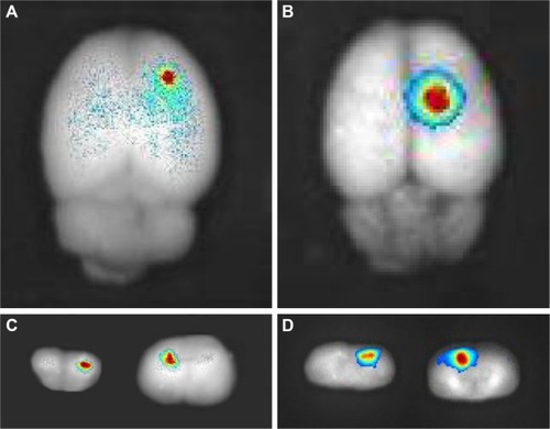
The antitumor effect of PAMAM-PEG-Tf/Trail was also reflected in the median survival time of rats bearing glioma (). After one treatment course, the median survival time of PAMAM-PEG-Tf/Trail-treated rats (28.5 days) was longer than that of rats treated with PAMAM-PEG/hTRAIL (25.5 days), Temozolomide (24.5 days) PAMAM-PEG-Tf/pEGFP (19 days), or saline (17 days).
Figure 8 Therapeutic effect evaluation.
Abbreviations: PAMAM, polyamidoamine dendrimer; PEG, polyethyleneglycol; pEGFP, plasmid encoding enhanced green fluorescence protein; MRI, magnetic resonance imaging; Tf, transferrin.
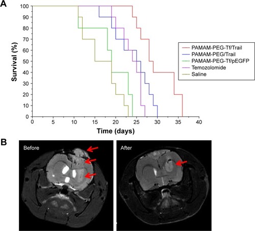
The anti-tumor effect of PAMAM-PEG-Tf/Trail was also confirmed by MRI imaging (). Tumor volume of PAMAM-PEG-Tf/hTRAIL-treated mice significantly decreased, which then prolonged the median survival time.
Discussion
Glioma is the most common malignant tumor of the central nervous system. Patients with malignant glioma have a poor prognosis due to the resistance to radiotherapy, chemotherapy, or immunotherapy.Citation31 The feature of invasive growing of glioma made it impossible for surgical resection. Gene therapy of Trail was proven to be a novel one.Citation32–Citation34 We had successfully synthesized the targeting dendrimer PAMAM-PEG-Tf for efficient gene delivery into brain.Citation26 It was demonstrated that Tf receptors expressed on the BBB and especially on tumor cells.Citation35,Citation36 Thus, this system was introduced to facilitate Trail crossing the BBB and targeting to the tumor.
A fundamental requirement for successful gene delivery was that dendrimer should be able to condense DNA efficiently to form NPs at a rational ratio. NPs should possess positive charge to enter the negatively charged cell membrane easily. At the weight ratio of PAMAM to DNA (3:1), PAMAM-PEG-Tf and DNA could form stable NPs with suitable size for enhanced permeability and retention (EPR) effect. The following cellular uptake and in vitro gene expression confirmed that PAMAM-PEG-Tf/DNA could enter the cells efficiently and yield high GFP expression. Theologically, the high PAMAM ratio could result in more stable NPs. However, cytotoxicity increased along with the PAMAM concentration due to the presence of surface amine groups.Citation37–Citation40 In the dendrimer design strategy, PEG could not only serve as a linker between PAMAM and Tf, but also reduce the toxicity by consuming some surface amines groups. Furthermore, PEG could provide a protective shell to avoid the reticulo-endothelial system, thus improving the half-life of NPs. Taken these together, 3:1 of PAMAM to DNA was optimized with tolerable toxicity and applied in the whole studies.
The glioma-targeting efficiency was also evaluated at the in vivo level. The fluorescent imaging showed that PAMAM-PEG-Tf/DNA NPs were more concentrated in glioma-bearing model than PAMAM-PEG/DNA NPs. PAMAM-PEG/DNA NPs could accumulate in tumor via EPR effects.Citation41 Whereas, PAMAM-PEG-Tf/DNA NPs could also accumulate in tumor via the receptor-mediated mechanism. The result of the following in vivo gene expression study was consistent with the aforementioned results. Tf-modified NPs expressed more GFP in tumor. The defective vascular architecture near the tumor margin was created due to the rapid vascularization necessary to serve fast-growing and invasive glioma. Thus, GFP expression of unmodified NPs, which was affected only by EPR effect, was found near the tumor margin. The distribution of Tf-modified NPs was affected by both EPR effect and Tf receptor. Thus, GFP expression can be also found in the inner tumor region. This demonstrated the targeting efficacy and superiority of PAMAM-PEG-Tf. The following investigation provided more evidence of this hypothesis. TUNEL assay results had the same trend with GFP expression that Tf-modified NPs had more apoptosis cells in both margin and central regions. Temozolomide, a commercial superiority drug for glioma, was selected as a positive control. It inactivated O6-alkylguanine-DNA alkyltransferase activity to make the DNA mismatched and lead to cytotoxicity, which was effective especially for cell division.Citation42 This was coincidental with the fact that Temozolomide treatment-induced apoptosis was only found in the margin region where the cells were proliferated rapidly. This difference of antitumor outcome between NP-based gene therapy and Temozolomide-based chemotherapy could provide us a new way to treat glioma more efficiently.
Conclusion
In conclusion, this study showed the feasibility of systemic administration of PAMAM-PEG-Tf/Trail for gene therapy of glioma. The current outcomes encourage further investigations into the application of gene therapy against tumor.
Acknowledgments
The authors would like to thank Mr XueFeng Yang for his technical support. This project has been funded in part with funds from the: Natural Science Foundation of Zhejiang Province (No. LY15H080002); Science and Technology Project of Zhejiang Province Department of Science and Technology (No. 2013C33230); Medical Science and Technology project of Zhejiang Province (No. 2012KYB088); Medical Science and Technology project of Zhejiang Province (No. 201337756); and Research project of Zhejiang education department (No. Y201328079).
Disclosure
The authors report no conflicts of interest in this work.
References
- SiegelinMDGaiserTHabelADaidzein overcomes TRAIL-resistance in malignant glioma cells by modulating the expression of the intrinsic apoptotic inhibitor, bcl-2Neurosci Lett2009454322322819429088
- RichJNBignerDDDevelopment of novel targeted therapies in the treatment of malignant gliomaNat Rev Drug Discov20043543044615136790
- GieseAGlioma invasion–pattern of dissemination by mechanisms of invasion and surgical intervention, pattern of gene expression and its regulatory control by tumorsuppressor p53 and proto-oncogene ETS-1Acta Neurochir Suppl20038815316214531573
- LefrancFBrotchiJKissRPossible future issues in the treatment of glioblastomas: special emphasis on cell migration and the resistance of migrating glioblastoma cells to apoptosisJ Clin Oncol200523102411242215800333
- DeAngelisLMBenefits of adjuvant chemotherapy in high-grade gliomasSemin Oncol2003306 Suppl 19151814765379
- OldenhuisCNStegehuisJHWalenkampAMTargeting TRAIL death receptorsCurr Opin Pharmacol20088443343918625341
- RiegerJNaumannUGlaserTAPO2 ligand: a novel lethal weapon against malignant glioma?FEBS Lett199842711241289613612
- KeaneMMEttenbergSANauMMChemotherapy augments TRAIL-induced apoptosis in breast cell linesCancer Res19995937347419973225
- PanGO’RourkeKChinnaiyanAMThe receptor for the cytotoxic ligand TRAILScience199727653091111139082980
- HawkinsCJTRAIL and malignant gliomaVitam Horm20046742745215110189
- RothWIsenmannSNaumannULocoregional Apo2L/TRAIL eradicates intracranial human malignant glioma xenografts in athymic mice in the absence of neurotoxicityBiochem Biophys Res Commun1999265247948310558893
- FukumuraDXuLChenYHypoxia and acidosis independently up-regulate vascular endothelial growth factor transcription in brain tumors in vivoCancer Res200161166020602411507045
- HobbsSKMonskyWLYuanFRegulation of transport pathways in tumor vessels: role of tumor type and microenvironmentProc Natl Acad Sci U S A1998958460746129539785
- MonskyWLFukumuraDGohongiTAugmentation of transvascular transport of macromolecules and nanoparticles in tumors using vascular endothelial growth factorCancer Res199959164129413510463618
- PardridgeWGene targeting technology and gene therapy of the brainDrug Discov Today20016312512611165183
- PardridgeWMDrug and gene delivery to the brain: the vascular routeNeuron200236455555812441045
- NewtonHBAdvances in strategies to improve drug delivery to brain tumorsExpert Rev Neurother20066101495150917078789
- GroothuisDRThe blood-brain and blood-tumor barriers: a review of strategies for increasing drug deliveryNeuro Oncol200021455911302254
- QianZMLiHSunHTargeted drug delivery via the transferrin receptor-mediated endocytosis pathwayPharmacol Rev200254456158712429868
- HatakeyamaHAkitaHMaruyamaKFactors governing the in vivo tissue uptake of transferrin-coupled polyethylene glycol liposomes in vivoInt J Pharm20042811–2253315288340
- PenichetMLKangYSPardridgeWMAn antibody-avidin fusion protein specific for the transferrin receptor serves as a delivery vehicle for effective brain targeting: initial applications in anti-HIV antisense drug delivery to the brainJ Immunol199916384421442610510383
- JefferiesWABrandonMRHuntSVTransferrin receptor on endothelium of brain capillariesNature198431259901621636095085
- TortorellaSKaragiannisTCTransferrin receptor-mediated endocytosis: a useful target for cancer therapyJ Membr Biol2014247429130724573305
- DixitSNovakTMillerKTransferrin receptor-targeted theranostic gold nanoparticles for photosensitizer delivery in brain tumorsNanoscale2015751782179025519743
- BielinskaAUChenCJohnsonJDNA complexing with polyamidoamine dendrimers: implications for transfectionBioconjug Chem199910584385010502352
- HuangRQQuYHKeWLEfficient gene delivery targeted to the brain using a transferrin-conjugated polyethyleneglycol-modified polyamidoamine dendrimerFASEB J20072141117112517218540
- PangZGaoHYuYEnhanced intracellular delivery and chemotherapy for glioma rats by transferrin-conjugated biodegradable polymersomes loaded with doxorubicinBioconjug Chem20112261171118021528923
- LvQLiLMHanMCharacteristics of sequential targeting of brain glioma for transferrin-modified cisplatin liposomeInt J Pharm20134441–21923347891
- ZhanCGuBXieCCyclic RGD conjugated poly(ethylene glycol)-co-poly(lactic acid) micelle enhances paclitaxel anti-glioblastoma effectJ Control Release2010143113614220056123
- LiJGuoYKuangYCholine transporter-targeting and co-delivery system for glioma therapyBiomaterials201334369142914823993342
- AvgeropoulosNGBatchelorTTNew treatment strategies for malignant gliomasOncologist19994320922410394589
- LuWSunQWanJCationic albumin-conjugated pegylated nanoparticles allow gene delivery into brain tumors via intravenous administrationCancer Res20066624118781188717178885
- HanLHuangRLiJPlasmid pORF-hTRAIL and doxorubicin co-delivery targeting to tumor using peptide-conjugated polyamidoamine dendrimerBiomaterials20113241242125220971503
- HuangSLiJHanLDual targeting effect of Angiopep-2-modified, DNA-loaded nanoparticles for gliomaBiomaterials201132286832683821700333
- MohsenikiaMAlizadehAMKhodayariSThe protective and therapeutic effects of alpha-solanine on mice breast cancerEur J Pharmacol20137181–31924051269
- TortorellaSKaragiannisTCThe significance of transferrin receptors in oncology: the development of functional nano-based drug delivery systemsCurr Drug Deliv201411442744324387131
- NahaPCDavorenMLyngFMReactive oxygen species (ROS) induced cytokine production and cytotoxicity of PAMAM dendrimers in J774A.1 cellsToxicol Appl Pharmacol20102461–2919920420846
- NahaPCDavorenMCaseyAAn ecotoxicological study of poly(amidoamine) dendrimers-toward quantitative structure activity relationshipsEnviron Sci Technol200943176864686919764261
- NahaPCByrneHJGeneration of intracellular reactive oxygen species and genotoxicity effect to exposure of nanosized polyamidoamine (PAMAM) dendrimers in PLHC-1 cells in vitroAquat Toxicol2013132–1336172
- MaherMANahaPCMukherjeeSPNumerical simulations of in vitro nanoparticle toxicity – the case of poly(amido amine) dendrimersToxicol In Vitro20142881449146025151936
- Brannon-PeppasLBlanchetteJONanoparticle and targeted systems for cancer therapyAdv Drug Deliv Rev200456111649165915350294
- TolcherAWGersonSLDenisLMarked inactivation of O6-alkylguanine-DNA alkyltransferase activity with protracted temozolomide schedulesBr J Cancer20038871004101112671695