
Abstract
Background
Intestinal ischemia-reperfusion (II/R) injury is a common clinical complication associated with high mortality, for which microRNA (miRNA) drives potentially its pathophysiological progression. MiRNAs regulate different messenger RNAs (mRNAs). However, the regulatory network between miRNAs and mRNAs in intestinal ischemia-reperfusion injury is elusive.
Methods
We analyzed the different expression of mRNAs and miRNAs in intestinal tissues from patients from three groups (arterial group (group A), venous group (group V), control group (group C)). Common differentially expressed (Co-DE) miRNAs and differentially expressed mRNAs were acquired via concerned analyses among the three groups. Co-DE mRNAs were shared parts of target mRNAs and differentially expression mRNAs. Cytoscape was employed to construct the regulatory network between miRNAs and mRNAs. Gene Ontology (GO) analysis and the Kyoto Encyclopedia of Genes and Genomes (KEGG) pathway depicted the functions and potential pathway associated with Co-DE mRNAs. Using the STRING and Cytoscape, we found critical mRNAs in the protein–protein interaction (PPI) network.
Results
The miRNA-mRNA network comprised 8 Co-DE miRNAs and 140 Co-DE mRNAs. Of note, 140 Co-DE mRNAs were targets of these 8 miRNAs, and their roles were established through the functional exploration via GO analysis and KEGG analysis. PPI network and Cytoscape revealed COL1A2, THY1, IL10, MMP2, SERPINH1, COL3A1, COL14A1, and P4HA1 as the top 8 key mRNAs.
Conclusion
This study has demonstrated a miRNA-mRNA regulatory network in intestinal ischemia-reperfusion injury, and explored the key mRNAs and their potential functions. These findings could provide new insight into prognostic markers and therapeutic targets for patients with intestinal ischemia-reperfusion injury in clinical practice.
Introduction
Intestinal ischemia-reperfusion (II/R) injury is a severe clinical complication common in the Intensive Care Unit (ICU). It is associated with high morbidity and mortality.Citation1 Usually, this problem is followed by various causes, including sepsis, shock, trauma, and so on.Citation2 Intestinal ischemia-reperfusion injury destroys intestinal tissue and impairs the function of the intestinal barrier. These events induce multiple organ dysfunction syndrome (MODS) or systemic inflammatory response syndrome (SIRS).Citation3,Citation4 The pathophysiological process of intestinal ischemia-reperfusion injury is complex, and different molecule mechanisms have been verified in this process.Citation5,Citation6 Ischemia injury and reperfusion injury are the two stages of intestinal ischemia-reperfusion injury.Citation7 The former is mainly caused by hypoxemia and results in the accumulation of much hypoxanthine in cells. During reperfusion, hypoxanthine reacts with oxygen molecules re-entering the cells to generate reactive oxygen species (ROS), which could make further damage on intestine.Citation8 In addition, intestinal ischemia causes the dysfunction of the intestinal barrier and activates inflammatory cells that release oxygen radicals.Citation9,Citation10 Compared with ischemia, the subsequent refusion usually leads to more terrible damage.Citation11 The severity of reperfusion injury is related to ischemia injury. Therefore, reducing ischemia injury is the key step to effective alleviation intestinal ischemia-reperfusion injury.
Various microRNAs (miRNAs), mRNAs, genes and proteins, the basic components of cells, contribute to the development of the intestinal ischemia-reperfusion injury. As a type of small non-coding RNA, miRNA regulates the expression of genes by accelerating mRNA degradation or inhibiting mRNA translation.Citation12,Citation13 Studies have confirmed that miRNA mediates the progression of intestinal ischemia-reperfusion injury. Nurr1 is associated with the regeneration of the intestinal epithelial and the function of the intestinal barrier. When miR-381-3p targets the Nurr1mRNA, intestinal epithelial proliferation activity decreased and the intestinal barrier function was impaired, which could aggravate the intestinal ischemia-reperfusion injury.Citation14 MiR-665-3p inhibits autophagy effects by targeting ATG4B, therefore, it can downregulate the inflammation and apoptosis in intestinal ischemia-reperfusion injury.Citation15 Caspase-3 expression is related to cell apoptosis in intestinal ischemia-reperfusion injury, and miR-378 and miR-182 both regulate Caspase-3 to exert a protective effect against this injury.Citation16,Citation17 Also, miR-21 potentially regulates the function of the intestinal barrier which is an integral part of intestinal ischemia-reperfusion injury.Citation18 However, further research is needed to explore the mechanism of miRNAs in II/R injury. MiRNA-mRNA interactions form a competitive endogenous RNA (ceRNA) regulatory network, widely used in pathophysiology and treatment of diseases.Citation19–Citation21
In this article, we tested the expression of mRNAs and miRNAs through transcriptome sequencing and small RNA sequencing in patients with intestinal ischemia-reperfusion injury. Then, GO analysis and KEGG analysis were applied to analyze mRNA and construct a miRNA-mRNA regulatory network. We aimed to identify biomarkers and new therapeutic targets for intestinal ischemia-reperfusion injury.
Patients and Methods
Patients and Sample Collection
We selected 13 patients (8 patients were with intestinal ischemia-reperfusion injury patients, and 5 patients without intestinal ischemia-reperfusion injury, all patients who underwent surgery were over 18 years old and were clearly diagnosed with intestinal injury during the surgical procedure in a medical ICU at Shanghai Zhongshan Hospital Affiliated to Fudan university) from the Intensive Care Unit (ICU) in Zhongshan Hospital (Shanghai, China), between July 2020 and March 2021. Intestinal epithelial tissue was collected from patients with intestinal ischemia-reperfusion injury (n=8) and normal control patients (group C, n=5) for follow-up tests. Patients with intestinal ischemia were categorized into the arterial group (Group A, n=5) and the venous group (Group V, n=3). Arterial group is defined as arterial ischemic intestinal injury caused by mesenteric artery embolism or thrombosis during the surgery. Venous group is defined as intestinal injury caused by venous congestion or incarceration leading to impaired blood circulation during the surgery. Intraoperatively resected intestinal specimens from patients in the enrolled arterial, venous, and control groups were excised in whole layers of approximately 1 cm, immediately transferred in a liquid nitrogen tank, and placed in a −80°C refrigerator for storage. All samples were obtained in accordance with the hospital’s regulations and the Ethics Committee of Shanghai Medical College of Fudan University approved the study. All patients signed the corresponding informed consent. This study was conducted in accordance with the Declaration of Helsinki.
RNA Isolation, Library Preparation and RNA Sequencing
The mRNA which had polyA enrichment was enriched by Oligo(dT), and RNA was fragmented using related RNA fragmentation reagent. The cDNA was synthesized by using random hexamer-primer and dNTPs. The qualified double-strand DNA library was transformed into a single-stranded circular DNA library through DNA-denaturation and circularization. Small RNA was selected from the total RNA, and then the 3ʹ end of the small RNA fragment was ligated by the 5-adenylated and 3-blocked adaptor. Unique molecular identifiers (UMI) labeled Primer was added into the 3ʹ end of the small RNA fragment and the 5ʹ end of the small RNA fragment was ligated. Single stranded cDNA was synthesized and amplified by PCR. The PCR products in the range of 110–130bp was isolated by PAGE electrophoresis. A highly efficient library was built. The Agilent 2100 Bioanalyzer was used to establish the quality of libraries. Libraries were sequenced on the DNBseq platform by Beijing Genomics Institute (BGI), China.
Analysis of Common Differentially Expressed (Co-DE) miRNAs
Differentially expressed (DE) miRNAs in intestinal ischemia-reperfusion injury from group C vs group A, group C vs group V were screened and analyzed: |log2 fold-change (log2FC)| ≥1 and p < 0.05 were set as the screening criterion. Co-DE miRNAs were DE miRNAs found in both comparison groups. The ggplot2 package in R software was adopted to generate volcano maps and heatmaps of DE miRNAs. Co-DE miRNAs were listed in the two comparison groups.
Predicting Target Genes of Co-DE miRNAs
Online databases (miRDB, miRWalk and TargetScan) were used to predict the potential targets of Co-DE miRNAs. To improve the accuracy of results, each target gene was listed in the three databases.
Analysis of Common Differentially Expressed (Co-DE) mRNAs
Differentially expressed (DE) mRNAs in intestinal ischemia-reperfusion injury from group A vs group C and group V vs group C were screened and analyzed. The screening criterion was |log2 fold-change (log2FC)| ≥1 and p-value < 0.05. mRNAs found in both DE mRNAs and target genes of Co-DE miRNAs were described as Co-DE mRNAs.
Constructing Co-DE miRNAs – Co-DE mRNAs Regulatory Network and Analyzing Biological Function of Co-DE mRNAs
Based on miRNAs and their target mRNAs, we constructed the network of Co-DE miRNA and Co-DE mRNA via Cytoscape (version 3.8.0). Data for Co-DE mRNAs in intestinal ischemia-reperfusion injury were subjected to Gene Ontology (GO) and Kyoto Encyclopedia of Genes and Genomes (KEGG) analysis to explore the function of mRNAs using package “clusterProfiler”, “AnnotationHub” and “org.Hs.eg.db”. Due to the small number of screened mRNA, we chose p-value < 0.05 as the screening criterion.
Constructing the Protein–Protein Interaction (PPI) Network
We acquired data of Co-DE mRNAs and constructed the PPI network via STRING database (https://string-db.org/). This allowed us to further understand interactions among Co-DE mRNAs. The following criterion was used: (1) Homo sapiens; (2) medium confidence 0.400. After that, the top eight Co-DE mRNAs were selected.
Results
Raw Sequence Reads and Work Flow
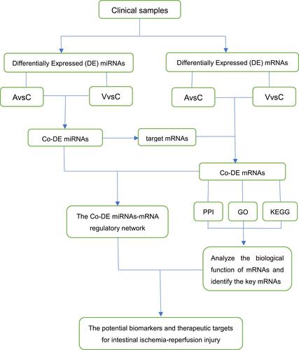
In RNA sequence, total clean reads were 118.99, 119.04, 47 million and clean reads ratio was over 99.15% from group A, C, and V, respectively (Table S1). In small RNA sequence, the proportion of clean tag was over 93.7% (Table S2). All data from intestinal ischemia-reperfusion injury were disposed and analyzed according to the work flow ().
Figure 1 The workflow of study.
Analysis of Differentially Expressed mRNA and miRNA in Intestinal Ischemia-Reperfusion Injury
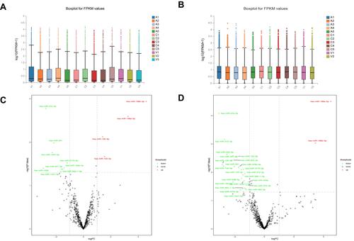
A total of 24,237 mRNAs and 3280 miRNAs were identified in three groups. The boxplots ( and ) revealed no statistical difference in the mRNA and miRNA distributions in the samples. Furthermore, 13 differentially expressed (DE) miRNAs were identified from group C vs group V, whereas 27 DE miRNAs were identified from group C vs group A ( and , Tables S3 and S4). According to the screening criterion (|log2FC|≥1, p-value < 0.05), there were 2902 DE mRNAs (1309 upregulated and 1593 downregulated, Table S5) and 931 DE mRNAs (489 upregulated and 442 downregulated, Table S6) in group C vs group A and group C vs group V, respectively.
Figure 2 Differentially expressed mRNAs and miRNAs in intestinal tissues in three groups. (A) A box plot of miRNAs in all samples. (B) A box plot of mRNAs in all samples. (C) A volcano plot of differentially expressed miRNAs from group C vs group V. (D) A volcano plot of differentially expressed miRNAs from group C vs group A.
Construction of ceRNA Regulatory Network
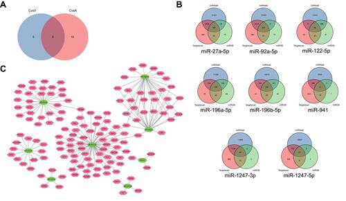
We found 8 common differentially expressed miRNAs (Co-DE miRNAs) (), including 7 consistent Co-DE miRNAs (2 upregulated and 5 downregulated) and 1 inconsistent Co-DE miRNAs (miR-122-5p, upregulated in group C vs group V and downregulated in group C vs group A). Total target mRNAs of Co-DE miRNAs were identified via online databases (). We selected 140 Co-DE mRNAs through the interaction analysis with target mRNAs (Table S7) and DE mRNAs. Finally, the regulatory network of Co-DE miRNAs and Co-DE mRNAs was constructed ().
Figure 3 The Co-DE miRNA-mRNA regulatory network. (A) A Venn plot of common differentially expressed miRNAs. (B) Venn plots of Co-DE mRNAs. (C) The constructed miRNA-mRNA regulatory network (the green ovals and pink polygons represent miRNAs and mRNAs respectively.).
GO and KEGG Analysis of Co-DE mRNAs
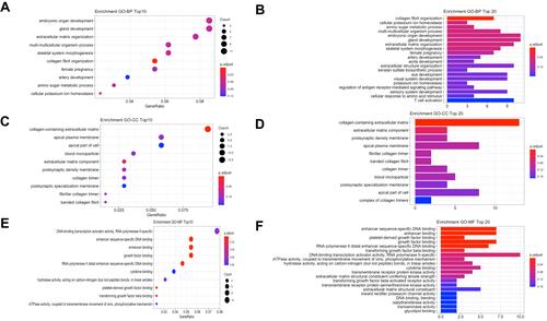
Following GO and KEGG analysis, 140 Co-DE mRNAs were used to establish the potential function of Co-DE miRNAs. The top 10 results from the GO biological process, GO cellular component and GO molecular function were presented in bubble charts (, and ), and the top 20 results were shown in bar charts (, and ). The top 20 results from KEGG analysis were presented in both bubble chart and bar chart ( and ).
Figure 4 Functional enrichment analysis of common differentially expressed mRNAs. (A) A dotplot of GO biological process (top 10 results). (B) A barplot of GO biological process (top 20 results). (C) A dotplot of GO cellular component (top 10 results). (D) A barplot of GO cellular component (top 20 results). (E) A dotplot of GO molecular function (top 10 results). (F) A barplot of GO molecular function (top 20 results). (G) A dotplot of KEGG analysis (top 20 results). (H) A barplot of KEGG analysis (top 20 results).
Construction of PPI Network and Identification of Hub mRNAs
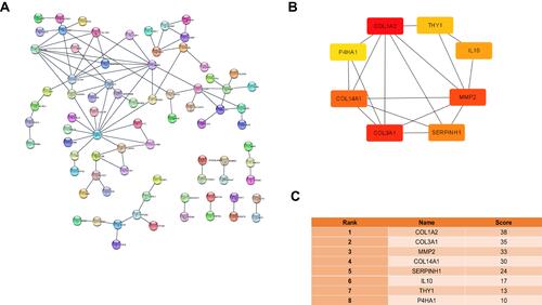
A total of 79 mRNAs from 140 Co-DE mRNAs were identified in the PPI network with 95 edges connected to each other (). The top 8 important mRNAs were identified via the cytoHubba in Cytoscape ( and ). They included Thy-1 cell surface antigen (THY1), collagen type I alpha 2 chain (COL1A2), collagen type XIV alpha 1 chain (COL14A1), prolyl 4-hydroxylase subunit alpha 1 (P4HA1), interleukin 10 (IL 10), collagen type III alpha 1 chain (COL3A1), matrix metalloproteinase-2 (MMP2) and serpin peptidase inhibitor, clade H, member 1 (SERPINH1).
Figure 5 PPI Network and Hub mRNAs. (A) PPI network of 79 mRNAs from all Common differentially expressed mRNAs. (B) The network of the top 8 key mRNAs. (C) The score and rank of key mRNAs in the PPI network.
Discussion
As a predominant high-risk disease, intestinal ischemia-reperfusion injury is associated with high mortality and a complex pathophysiological process in the Intensive Care Unit (ICU).Citation22 Several severe conditions, including shock, thrombus and others, are the predisposing factors for this disease.Citation23 Because of the damage to the intestinal barrier, patients are more prone to the translation of bacterial, septicemia, and multi-organ failure.Citation24 Previously, there were several studies related to the role of miRNA in intestinal ischemia-reperfusion injury.Citation14–Citation16 However, the pathophysiological mechanism of this disease remains unclear.
Through transcriptome sequencing, we explored the expression profiles of mRNAs and miRNAs in the intestinal tissues of group A, group C, and group V. Moreover, the role of Co-DE miRNAs and the biological function of Co-DE mRNAs were established via GO and KEGG analyses. Our study aimed to uncover biomarkers and potential therapeutic targets for intestinal ischemia-reperfusion injury.
With the technological advancement, non-coding RNAs (ncRNAs), such as microRNA (miRNA), long non-coding RNA (lncRNA), and circRNA, have been discovered. More studies on the regulation of genes are geared towards ncRNAs.Citation25 According to previous research, all eight Co-DE miRNAs could be speculated as the potential regulatory targets in intestinal ischemia-reperfusion injury. As an inconsistent Co-DE miRNA, the expression of miR-122-5p varied in intestinal ischemia-reperfusion injury and ischemia-reperfusion induced arrhythmias.Citation26 MiR-27a-5p had been associated with a decreased level of Bach1 mRNA, which reduced apoptosis in liver ischemia-reperfusion injury.Citation27 Elsewhere, miR-1247-3p alleviated the damage of myocardial ischemia-reperfusion by targeting both BCL2L11 and caspase-2.Citation28 In addition, miR-1247-3p reduced apoptosis of cerebral neurons and could exert protective effects against brain stroke.Citation29 Although available research indicates that other Co-DE miRNAs are not directly related to ischemia-reperfusion injury, they all are associated with apoptosis, a key player in intestinal ischemia-reperfusion injury.Citation30–Citation33 These Co-DE miRNAs may be associated with the occurrence and progression of intestinal ischemia-reperfusion injury, and are potential therapeutic targets.
Through GO and KEGG analysis, identified the main biological function of Co-DE mRNAs. There are several signaling pathways involved in ischemia-reperfusion injury, including Wnt signaling pathway,Citation34 PI3K/Akt signaling pathway,Citation35 JAK/STAT signaling pathway,Citation32 and cAMP signaling pathwayCitation36 among others. The result of KEGG analysis verified the association of Co-DE mRNAs with the above signaling pathways, such as cAMP pathway and PI3K/Akt pathway. Using the cytoHubba, we revealed the top eight key mRNAs, including Thy-1 cell surface antigen (THY1), collagen type I alpha 2 chain (COL1A2), collagen type XIV alpha 1 chain (COL14A1), prolyl 4-hydroxylase subunit alpha 1 (P4HA1), interleukin 10 (IL 10), collagen type III alpha 1 chain (COL3A1), matrix metalloproteinase-2 (MMP2) and serpin peptidase inhibitor, clade H, member 1 (SERPINH1). The activity of Wnt signaling pathway was related to whether the cells expressed THY1 or not.Citation37 Existing reports show that MAPK pathway exerts a negative regulatory effect on the expression of COL1A2. Of note, MAPK pathway plays a key role in the critical phase of ischemia/reperfusion injury,Citation38,Citation39 and regulates the expression of P4HA1.Citation40 Studies have revealed that IL10 mediates the activity of MAPK pathway, AMPK pathway, and JAK/STAT pathway,Citation41–Citation43 and COL3A1 was identified as the regulatory factor of MAPK pathway.Citation44 MMP2, as a kind of downstream gene, was found to be regulated by MAPK pathway, Wnt pathway, and JAK/STAT pathway, which were involved in the pathophysiological process of ischemia-reperfusion injury.Citation45–Citation47 Thus, all the top 8 mRNAs may play important roles in the prediction and treatment of this disease, an area that warrants further research.
There are a few limitations in our study. Firstly, because of the small number of patients, the link between the clinical information of samples and the ceRNA regulatory network could not be analyzed. Secondly, our findings are mainly based on bioinformatics analysis, further mechanistic analysis of cells and animals did not be covered. In future studies, the collection of clinical samples should be increased. Besides, we will further identify and confirm specific functional miRNA and mRNA through research on mechanism.
In conclusion, this work explored the expression profiles of mRNAs and miRNAs, which has allowed for the construction of a ceRNA regulatory network for intestinal ischemia-reperfusion injury. Further, the potential function of Co-DE miRNAs and mRNAs has been established, through functional enrichment analysis on these mRNAs via GO and KEGG analysis. The findings provide a new view on identifying biomarkers and therapeutic targets for intestinal ischemia-reperfusion injury.
Data Available
The data for this study has been deposited in the European Nucleotide Archive (ENA) at EMBL-EBI under accession number PRJEB45362 (https://www.ebi.ac.uk/ena/browser/view/PRJEB45362).
Ethics Approval and Informed Consent
All research studies on humans (individuals, samples or data) include a statement on ethics approval and consent.
Disclosure
The authors report no conflicts of interest in this work.
Additional information
Funding
References
- Chen F, Wang D, Li X, Wang H. Molecular mechanisms underlying intestinal ischemia/reperfusion injury: bioinformatics analysis and in vivo validation. Med Sci Monit. 2020;26:e927476. doi:10.12659/MSM.927476
- Corcos O, Nuzzo A. Gastro-intestinal vascular emergencies. Best Pract Res Clin Gastroenterol. 2013;27(5):709–725. doi:10.1016/j.bpg.2013.08.006
- Brath E, Nemeth N, Kiss F, et al. Changes of local and systemic hemorheological properties in intestinal ischemia-reperfusion injury in the rat model. Microsurgery. 2010;30(4):321–326. doi:10.1002/micr.20707
- Mester A, Magyar Z, Sogor V, et al. Intestinal ischemia-reperfusion leads to early systemic micro-rheological and multiorgan microcirculatory alterations in the rat. Clin Hemorheol Microcirc. 2018;68(1):35–44. doi:10.3233/CH-170278
- Berlanga J, Prats P, Remirez D, et al. Prophylactic use of epidermal growth factor reduces ischemia/reperfusion intestinal damage. Am J Pathol. 2002;161(2):373–379. doi:10.1016/S0002-9440(10)64192-2
- Karkkainen JM, Acosta S. Acute mesenteric ischemia (part I) - Incidence, etiologies, and how to improve early diagnosis. Best Pract Res Clin Gastroenterol. 2017;31(1):15–25. doi:10.1016/j.bpg.2016.10.018
- Nadatani Y, Watanabe T, Shimada S, Otani K, Tanigawa T, Fujiwara Y. Microbiome and intestinal ischemia/reperfusion injury. J Clin Biochem Nutr. 2018;63(1):26–32. doi:10.3164/jcbn.17-137
- Irani K. Oxidant signaling in vascular cell growth, death, and survival a review of the roles of reactive oxygen species in smooth muscle and endothelial cell mitogenic and apoptotic signaling. Circ Res. 2000;87(3):179–183. doi:10.1161/01.RES.87.3.179
- Vollmar B, Menger M. Intestinal ischemia/reperfusion: microcirculatory pathology and functional consequences. Langenbeck’s Arch Surg. 2011;396(1):13–29. doi:10.1007/s00423-010-0727-x
- Blum H, Summers J, Schnall M, et al. Acute intestinal ischemia studies by phosphorus nuclear magnetic resonance spectroscopy. Ann Surg. 1986;204(1):83–88. doi:10.1097/00000658-198607000-00012
- Kalogeris T, Baines C, Krenz M, Korthuis R. Cell biology of ischemia/reperfusion injury. Int Rev Cell Mol Biol. 2012;298:229–317.
- Bartel D. MicroRNAs: target recognition and regulatory functions. Cell. 2009;136(2):215–233. doi:10.1016/j.cell.2009.01.002
- Lee Y, Ahn C, Han J, et al. The nuclear RNase III Drosha initiates microRNA processing. Nature. 2003;425(6956):415–419. doi:10.1038/nature01957
- Liu L, Yao J, Li Z, et al. miR-381-3p knockdown improves intestinal epithelial proliferation and barrier function after intestinal ischemia/reperfusion injury by targeting nurr1. Cell Death Dis. 2018;9(3):411. doi:10.1038/s41419-018-0450-z
- Li Z, Wang G, Feng D, et al. Targeting the miR-665-3p-ATG4B-autophagy axis relieves inflammation and apoptosis in intestinal ischemia/reperfusion. Cell Death Dis. 2018;9(5):483. doi:10.1038/s41419-018-0518-9
- Li Y, Wen S, Yao X, et al. MicroRNA-378 protects against intestinal ischemia/reperfusion injury via a mechanism involving the inhibition of intestinal mucosal cell apoptosis. Cell Death Dis. 2017;8(10):e3127. doi:10.1038/cddis.2017.508
- Luo Y, Duan X, Bian L, Chen Z, Kuang L, Li Y. Ischemic preconditioning preventing downregulation of miR-182 protects intestine against ischemia/reperfusion injury by inhibiting apoptosis. Arch Med Res. 2019;50(5):241–248. doi:10.1016/j.arcmed.2019.08.013
- Zhang L, Zhang F, He D, Fan X, Shen J. MicroRNA-21 is upregulated during intestinal barrier dysfunction induced by ischemia reperfusion. Kaohsiung J Med Sci. 2018;34(10):556–563. doi:10.1016/j.kjms.2018.05.006
- Qi X, Zhang D, Wu N, Xiao J, Wang X, Ma W. ceRNA in cancer: possible functions and clinical implications. J Med Genet. 2015;52(10):710–718. doi:10.1136/jmedgenet-2015-103334
- Sun F, Liang W, Tang K, Hong M, Qian J. Profiling the lncRNA-miRNA-mRNA ceRNA network to reveal potential crosstalk between inflammatory bowel disease and colorectal cancer. PeerJ. 2019;7:e7451. doi:10.7717/peerj.7451
- He L, Chen Y, Hao S, Qian J. Uncovering novel landscape of cardiovascular diseases and therapeutic targets for cardioprotection via long noncoding RNA-miRNA-mRNA axes. Epigenomics. 2018;10(5):661–671. doi:10.2217/epi-2017-0176
- Liu S, He X, Zhang X, Zeng F, Wang F, Zhou X. Ischemic preconditioning-Induced SOCS-1 Protects rat intestinal ischemia reperfusion injury via degradation of TRAF6. Dig Dis Sci. 2017;62(1):105–114. doi:10.1007/s10620-016-4277-0
- Koike Y, Li B, Lee C, et al. The intestinal injury caused by ischemia-reperfusion is attenuated by amniotic fluid stem cells via the release of tumor necrosis factor-stimulated gene 6 protein. FASEB J. 2020;34(5):6824–6836. doi:10.1096/fj.201902892RR
- Li Y, Feng D, Wang Z, et al. Ischemia-induced ACSL4 activation contributes to ferroptosis-mediated tissue injury in intestinal ischemia/reperfusion. Cell Death Differ. 2019;26(11):2284–2299. doi:10.1038/s41418-019-0299-4
- Beermann J, Piccoli M, Viereck J, Thum T. Non-coding RNAs in development and disease: background, mechanisms, and therapeutic approaches. Physiol Rev. 2016;96(4):1297–1325. doi:10.1152/physrev.00041.2015
- Tang J, Gao H, Liu Y, et al. Network construction of aberrantly expressed miRNAs and their target mRNAs in ventricular myocardium with ischemia-reperfusion arrhythmias. J Cardiothorac Surg. 2020;15(1):216. doi:10.1186/s13019-020-01262-4
- Xing Y, Li J, Li S, et al. MiR-27a-5p regulates apoptosis of liver ischemia-reperfusion injury in mice by targeting Bach1. J Cell Biochem. 2018;119(12):10376–10383. doi:10.1002/jcb.27383
- Huang J, Huang Y, Feng Z, Guo W, Wang X, Liao Z. MiR-1247-3p protects rat cardiomyocytes against hypoxia/reoxygenation-induced injury via targeting BCL2L11 and caspase-2. J Recept Signal Transduct Res. 2021;41(1):6–14. doi:10.1080/10799893.2020.1783554
- Zhang R, Zhou W, Yu Z, et al. miR-1247-3p mediates apoptosis of cerebral neurons by targeting caspase-2 in stroke. Brain Res. 2019;1714:18–26. doi:10.1016/j.brainres.2019.02.020
- Zeng B, Li Y, Feng Y, et al. Downregulated miR-1247-5p associates with poor prognosis and facilitates tumor cell growth via DVL1/Wnt/β-catenin signaling in breast cancer. Biochem Biophys Res Commun. 2018;505(1):302–308. doi:10.1016/j.bbrc.2018.09.103
- Li S, Cheng W, Li Y, et al. Keap1-targeting microRNA-941 protects endometrial cells from oxygen and glucose deprivation-re-oxygenation via activation of Nrf2 signaling. Cell Commun Signaling. 2020;18(1):32. doi:10.1186/s12964-020-0526-0
- Wang L, Wei Y, Yan Y, et al. CircDOCK1 suppresses cell apoptosis via inhibition of miR‑196a‑5p by targeting BIRC3 in OSCC. Oncol Rep. 2018;39(3):951–966.
- Zhai H, Zhang X, Chen S, Fan M, Ma S, Sun X. RP5-1120P11.3 promotes hepatocellular carcinoma development via the miR-196b-5p-WIPF2 axis. Biochemistry and Cell Biology. 2020;98(2):238–248. doi:10.1139/bcb-2019-0053
- Liu D, Liu Y, Zheng X, Liu N. c-MYC-induced long noncoding RNA MEG3 aggravates kidney ischemia-reperfusion injury through activating mitophagy by upregulation of RTKN to trigger the Wnt/β-catenin pathway. Cell Death Dis. 2021;12(2):191. doi:10.1038/s41419-021-03466-5
- Li Y, Chen Z, Cai Y. Piperine protects against myocardial ischemia/reperfusion injury by activating the PI3K/AKT signaling pathway. Exp Ther Med. 2021;21(4):374. doi:10.3892/etm.2021.9805
- Liu Y, Zhang J, Zan J, Zhang F, Liu G, Wu A. Lidocaine improves cerebral ischemia-reperfusion injury in rats through cAMP/PKA signaling pathway. Exp Ther Med. 2020;20(1):495–499. doi:10.3892/etm.2020.8688
- Aulicino F, Theka I, Ombrato L, Lluis F, Cosma M. Temporal perturbation of the Wnt signaling pathway in the control of cell reprogramming is modulated by TCF1. Stem Cell Rep. 2014;2(5):707–720. doi:10.1016/j.stemcr.2014.04.001
- Okano K, Tsuruta Y, Yamashita T, Takano M, Echida Y, Nitta K. Suppression of renal fibrosis by galectin-1 in high glucose-treated renal epithelial cells. Exp Cell Res. 2010;316(19):3282–3291. doi:10.1016/j.yexcr.2010.08.015
- Jeong Y, Oh Y, Cho W, Lee B, Ma J. Anti-inflammatory effects of melandrii herba ethanol extract via inhibition of NF-κB and MAPK Signaling Pathways and Induction of HO-1 in RAW 264.7 Cells and Mouse Primary Macrophages. Molecules. 2016;21:6. doi:10.3390/molecules21060818
- Zhang K, Huang XZ, Li XN, et al. Interleukin 6 destabilizes atherosclerotic plaques by downregulating prolyl-4-hydroxylase alpha1 via a mitogen-activated protein kinase and c-Jun pathway. Arch Biochem Biophys. 2012;528(2):127–133. doi:10.1016/j.abb.2012.09.007
- Thompson D, Morrice N, Grant L, et al. Myeloid protein tyrosine phosphatase 1B (PTP1B) deficiency protects against atherosclerotic plaque formation in the ApoE(-/-) mouse model of atherosclerosis with alterations in IL10/AMPKalpha pathway. Mol Metab. 2017;6(8):845–853. doi:10.1016/j.molmet.2017.06.003
- Priya GB, Nagaleekar VK, Milton AAP, et al. Genome wide host gene expression analysis in mice experimentally infected with Pasteurella multocida. PLoS One. 2017;12(7):e0179420. doi:10.1371/journal.pone.0179420
- Qian Q, Wu C, Chen J, Wang W. Relationship between IL10 and PD-L1 in Liver hepatocellular carcinoma tissue and cell lines. Biomed Res Int. 2020;2020:8910183. doi:10.1155/2020/8910183
- Fernandez-Serra A, Moura DS, Sanchez-Izquierdo MD, et al. Prognostic Impact of let-7e MicroRNA and Its Target Genes in Localized High-Risk Intestinal GIST: a Spanish Group for Research on Sarcoma (GEIS) Study. Cancers. 2020;12:10. doi:10.3390/cancers12102979
- Zhao F, Zhou L, Liu J, et al. Construction of a vascularized bladder with autologous adipose-derived stromal vascular fraction cells combined with bladder acellular matrix via tissue engineering. J Tissue Eng. 2019;10:2041731419891256. doi:10.1177/2041731419891256
- Qin L, Liao L, Redmond A, et al. The AIB1 oncogene promotes breast cancer metastasis by activation of PEA3-mediated matrix metalloproteinase 2 (MMP2) and MMP9 expression. Mol Cell Biol. 2008;28(19):5937–5950. doi:10.1128/MCB.00579-08
- Ghosh A, Pechota A, Coleman D, Upchurch G, Eliason J. Cigarette smoke-induced MMP2 and MMP9 secretion from aortic vascular smooth cells is mediated via the Jak/Stat pathway. Hum Pathol. 2015;46(2):284–294. doi:10.1016/j.humpath.2014.11.003