Abstract
Intrahepatic cholangiocarcinomas (IHCCs) are morphologically and biologically similar to pancreatic ductal adenocarcinomas (PDACs), so newly identified PDAC-associated genes or proteins could provide clues for screening novel biomarkers for IHCC. In this study, the expression of three novel PDAC tumor markers (T-box transcription factor-4 [TBX4], heat shock protein-60 [HSP60], and Parkinson protein-7 [DJ-1]) identified in previous proteomic studies in IHCC tumors were immunohistochemically detected. The current study confirmed that three novel pancreatic cancer biomarkers TBX4, HSP60, and DJ-1 were also overexpressed in IHCC tumors, but with a relatively lower expression level than PDAC. No significant association was found between tumor marker expression and the clinicopathological characteristics of IHCC patients except that TBX4 expression correlated with tumor grades. Moreover, DJ-1 was demonstrated to be an independent prognostic factor for these patients. The current findings suggest that DJ-1 might play an important role in the malignant progression of IHCC, and its exact mechanism during IHCC progression deserves further investigation.
Introduction
Intrahepatic cholangiocarcinomas (IHCCs) arising from the epithelia of the intrahepatic bile duct rank second in the primary malignant liver tumors. The incidence and mortality of IHCCs have increased in recent years in China, and this tendency has also been observed in Western countries.Citation1 Due to the lack of tumor markers for early diagnosis and effective optional therapeutic approaches, the prognosis of IHCC patients is very poor. Therefore, there is a great need to mine novel tumor markers as diagnostic, prognostic, and therapeutic targets for IHCC patients.
IHCCs are morphologically and biologically similar to pancreatic ductal adenocarcinomas (PDACs), most of the tumor markers of PDAC show similar expression patterns in biliary carcinomas.Citation2 Proteomics technology has become a powerful and efficient means for mining candidate tumor biomarkers with altered expression patterns in human malignancies. The authors and other groups have recently used multiple proteomic platforms to identify and validate a panel of novel candidate protein tumor markers that are overexpressed in PDAC tissues.Citation3,Citation4 As relatively fewer biomarkers have been identified in IHCCs, it was speculated that these PDAC proteins could provide a source for screening novel tumor markers for IHCC.
In a previous study using two-dimensional electrophoresis-based proteomic tools, three candidate biomarkers (T-box transcription factor-4 [TBX4], heat shock protein-60 [HSP60], and Parkinson protein-7 [DJ-1]) for PDAC were identified.Citation3–Citation6 These three proteins were overexpressed in PDAC tumors compared with normal pancreas tissues or benign tumors; furthermore, their expression levels were demonstrated to be associated with clinicopathological traits such as tumor grades. In particular, high expression of TBX4 was also an independent prognostic factor for PDAC.Citation5 In this study, the expression of the above three novel PDAC-associated proteins in the IHCC tumors were immunohistochemically assessed in order to validate if these proteins are overexpressed in IHCCs. Their clinicopathological and prognostic significance were also evaluated.
Material and methods
Patients
A total of 72 consecutive IHCC patients who underwent standard curative resection at the Department of General Surgery of Zibo Central Hospital (Zibo, China) and Qilu Hospital (Jinan, China) from 1998 to 2007 were reviewed retrospectively. All the patients routinely received postoperative gemcitabine-based chemotherapy regimens, and no patient received preoperative chemotherapy and/or radiation. The overall survival time of IHCC patients was defined as the time from operation to cancer-caused death. The clinicopathological data of these patients are summarized in . An additional 55 patients with PDAC who underwent standard curative resection at the Department of General Surgery of Zibo Central Hospital and Qilu Hospital from 2005 to 2007 were also included in this study. The study was approved by the local research ethics committee at the authors’ institution. Informed written consents were obtained from all patients.
Table 1 Relationship between tumor markers and clinicopathological characteristics in intrahepatic cholangiocarcinomas
Immunohistochemistry
Immunohistochemical staining was performed on 4 μm formalin-fixed, paraffin-embedded archival tissue sections of 72 IHCC primary tumors, their paired adjacent noncancerous tissues, and 55 PDAC tumors. Briefly, deparaffinized and rehydrated sections were boiled in sodium citrate buffer (pH 6.0) to perform antigen retrieval. Endogenous peroxidase activity was blocked by incubating the slides in methanol containing 3% hydrogen peroxide. Sections were incubated overnight at 4°C with the primary antibodies – anti-TBX4, 1:200 (Abcam, Cambridge, MA, USA), anti-HSP60, 1:100 (Santa Cruz Biotechnology, Santa Cruz, CA, USA), and anti-DJ-1, 1:150 (Santa Cruz Biotechnology) – and subsequently developed with an avidin–biotin–peroxidase complex method according to the manufacturer’s recommendations (EnVision™ system; Dako Denmark A/S, Glostrup, Denmark). The specificity of antibodies used in this study had been validated in previous studies.Citation3–Citation6 Diaminobenzidine was used as a chromogen, and hematoxylin was used as a counterstain. Nonspecific immunoglobulin G was used as a negative control. The expression level of each tumor marker was measured as high expression when moderate or strong staining was present in more than 25% of the cancer cells, which is accordance with previously established criteria on PDACs.
Statistics
The association of tumor biomarkers with the clinicopathological characteristics of IHCC patients was evaluated with Chi-squared or Fisher’s exact test as appropriate. Overall survival in relation to TBX4, HSP60, and DJ-1 expression patterns and other clinicopathological characteristics was evaluated by Kaplan–Meier survival curve and log-rank test. Multivariable analyses were analyzed using a Cox proportional hazards model. Only statistically significant prognostic factors identified by univariate analysis were entered in the multivariate analysis. Statistical analyses were performed using SPSS® 16.0 software for Windows (IBM Corporation, Armonk, NY). The level of statistical significance was set at P < 0.05.
Results
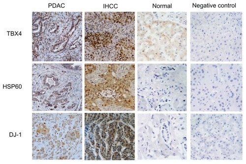
The expression of three recently identified PDAC markers (TBX4, HSP60, and DJ-1) in IHCC tissues was determined (). TBX4 showed mainly nuclear staining, and cytoplasmic staining was also frequently observed. Meanwhile, DJ-1 staining was mainly located in the cytoplasm, occasionally nuclear staining was also detected. HSP60 immunostaining was primarily in the cytoplasm and the membrane of neoplastic epithelium in IHCC tumors. The percentage of carcinomas demonstrating either focal or diffuse labeling with these markers varied greatly among IHCC samples. According to established criteria for immunostaining, 52.8%, 52.8%, and 54.2% of the IHCC tumors were defined as positive for TBX4, HSP60, and DJ-1, respectively. PDAC samples presented similar immunostaining patterns for TBX4, HSP60, and DJ-1 in IHCC tumors; however, all of these markers showed lower frequencies of expression in the IHCC samples in comparison to the PDAC samples (69.1% for TBX4, 83.6% for HSP60, and 76.3% for DJ-1). There were also significant differences in the levels of TBX4, HSP60, and DJ-1 expression between neoplastic and adjacent nonneoplastic intrahepatic duct epithelia (2.78% for TBX4, 6.94% for HSP60, and 2.78% for DJ-1) in IHCC tumors.
Figure 1 Representative images of the high expression of TBX4, HSP60, and DJ-1 in IHCC and PDAC tumors, and low or negative expression of them in the normal liver tissues (containing intrahepatic duct) and negative controls (IHCC tumors).
Because the IHCC tumors showed variable labeling with HSP60, DJ-1, and TBX4, their clinicopathological and prognostic significance were further evaluated. As seen in , no significant association was identified between tumor markers expression and the clinicopathological characteristics of IHCC patients except that a high level of TBX4 expression had a positive correlation with better differentiation types (grades 1–2). Kaplan–Meier analysis revealed a tendency towards shorter overall survival in patients with high DJ-1 expression, but not with TBX4 and HSP60 overexpression (). In this study, three factors including tumor size, lymph node metastasis, and DJ-1 expression were identified as significant prognostic factors by univariate analysis. Cox proportional hazard model analysis using these three factors confirmed that DJ-1 expression was an independent predictor for reduced overall survival of IHCC patients ().
Figure 2 Kaplan–Meier survival curves performed according to DJ-1 expression in IHCC patients.
Abbreviations: cum, cumulative; DJ-1, Parkinson protein-7; IHCC, intrahepatic cholangiocarcinoma.
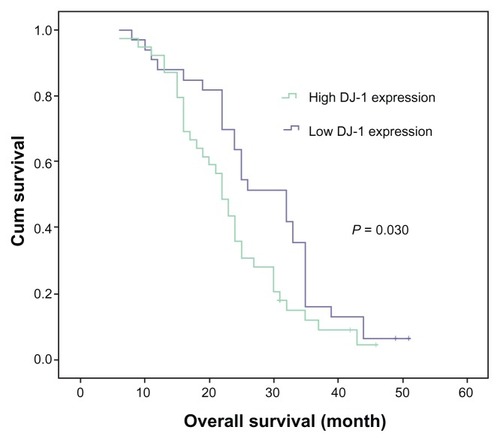
Table 2 Univariate and multivariate analysis of overall survival in patients with intrahepatic cholangiocarcinomas
Discussion
IHCCs are similar to PDACs based on the morphological and molecular backgrounds. A panel of tumors such as carbohydrate antigen-19-9, Dupan-2, carbohydrate antigen-50, Span-1, carcinoembryonic antigen, and pancreatic elastase-1 has been used in the diagnosis of both PDACs and IHCCs.Citation2 Compared with PDACs, less frequent studies focus on the mining of tumor markers in IHCC. Therefore, it is proposed that previously identified overexpressed genes or proteins in PDACs could provide clues for screening novel biomarkers for IHCC. For example, Swierczynski et al analyzed eight novel PDAC markers (fascin, mucin-4, 14-3-3σ, prostate stem cell antigen, topoisomerase-IIα, and cdc2/p34) in IHCC tumors.Citation7 The results of that study indicated that most PDAC markers show similar, yet not identical, expression patterns in biliary carcinomas, suggesting novel PDAC markers are potentially useful in developing diagnostic and therapeutic targets for tumors involving the biliary system.
The current study compared the performance of three novel PDAC markers TBX4, HSP60, and DJ-1 in IHCC samples. These PDAC markers were identified in previous proteomic studies, and have not been investigated in IHCC in previous studies. The results indicate that analogous to PDACs, IHCCs demonstrated overexpression of each of the markers examined, but their expression levels were relatively lower than those in PDACs. This trend is similar to that found in previous studies.
In a previous comparative proteomic study on pancreatic cancer and normal pancreas tissues, for the first time TBX4 and HSP60 were identified to be upregulated in PDACs.Citation3 TBX4 belongs to a transcription factor family characterized by a highly conserved DNA-binding motif (T-box) and plays a key role during organogenesis and pattern formation in both vertebrate and invertebrate embryos.Citation8,Citation9 HSP60 is a chaperonin protein involved in the folding and assembly of multiple proteins.Citation10 It was further validated that TBX4 was a differentiation-associated marker for PDAC, and low expression of TBX4 also predicted worse prognosis for patients with PDAC.Citation5 However, in this study, no association was observed between these two markers with the clinicopathological traits and prognosis of IHCC patients.
In another quantitative proteomics study on pancreatic juices, DJ-1 was identified to be overexpressed in PDAC tissue and pancreatic juice samples, and it was further confirmed that DJ-1 correlated with tumor differentiation in PDAC.Citation4 DJ-1 is a novel mitogen-dependent oncoprotein involved in Ras-related signal transduction pathway,Citation11 which has been confirmed to be upregulated in multiple cancers.Citation12,Citation13 In the current study, it was found that high DJ-1 expression level is an independent prognostic factor for IHCC patients. This finding suggests that DJ-1 might play an important role in the malignant progression of IHCC, and could be used as a potential candidate predictive factor for IHCC patients undergoing surgical resection.
Conclusion
In conclusion, the current study confirmed that three novel pancreatic cancer biomarkers TBX4, HSP60, and DJ-1 were also overexpressed in IHCC tumors, but with a relatively lower expression level than PDACs. DJ-1 might be associated with more progressive behaviors and worse prognosis for IHCC patients; its exact mechanism during IHCC progression deserves further investigation.
Disclosure
The authors report no conflicts of interest in this work.
References
- ShinHROhJKMasuyerEComparison of incidence of intrahepatic and extrahepatic cholangiocarcinoma – focus on East and South-Eastern AsiaAsian Pac J Cancer Prev20101151159116621198257
- TakezakoYOkusakaTUenoHIkedaMMorizaneCNajimaM(Tumor markers for pancreatic and biliary tract cancer)Gan To Kagaku Ryoho200431914431446 Japanese15446574
- QiTHanJCuiYZongMLiuXZhuBComparative proteomic analysis for the detection of biomarkers in pancreatic ductal adenocarcinomasJ Clin Pathol2008611495817412869
- TianMCuiYZSongGHProteomic analysis identifies MMP-9, DJ-1 and A1BG as overexpressed proteins in pancreatic juice from pancreatic ductal adenocarcinoma patientsBMC Cancer2008824118706098
- ZongMMengMLiLLow expression of TBX4 predicts poor prognosis in patients with stage II pancreatic ductal adenocarcinomaInt J Mol Sci20111284953496321954337
- CuiYTianMZongMProteomic analysis of pancreatic ductal adenocarcinoma compared with normal adjacent pancreatic tissue and pancreatic benign cystadenomaPancreatology200991–2899819077459
- SwierczynskiSLMaitraAAbrahamSCAnalysis of novel tumor markers in pancreatic and biliary carcinomas using tissue microarraysHum Pathol200435335736615017593
- AgulnikSIGarveyNHancockSEvolution of mouse T-box genes by tandem duplication and cluster dispersionGenetics199614412492548878690
- NaicheLAPapaioannouVELoss of Tbx4 blocks hindlimb development and affects vascularization and fusion of the allantoisDevelopment2003130122681269312736212
- RothmanJESchekmanRMolecular mechanism of protein folding in the cellCell2011146685185421907398
- van der BrugMPBlackintonJChandranJRNA binding activity of the recessive parkinsonism protein DJ-1 supports involvement in multiple cellular pathwaysProc Natl Acad Sci U S A200810529102441024918626009
- OdaMMakitaMIwayaKHigh levels of DJ-1 protein in nipple fluid of patients with breast cancerCancer Sci201210361172117622404125
- ChenYKangMLuWDJ-1, a novel biomarker and a selected target gene for overcoming chemoresistance in pancreatic cancerJ Cancer Res Clin Oncol201213891463147422526154