
Abstract
A 72-year-old woman was referred to our hospital with Stage IV non-small-cell lung cancer (NSCLC). Chest computed tomography revealed a mass in the upper lobe of the right lung, with pleural effusion. Cytologic examination identified adenocarcinoma cells in the right pleural effusion. Furthermore, both a deletion mutation in exon 19 and a threonine–methionine substitution mutation at position 790 in exon 20 (T790M) were detected in the epidermal growth factor receptors (EGFR) in the malignant cells. As systemic chemotherapy consisting of carboplatin and pemetrexed or erlotinib proved ineffective, docetaxel monotherapy was initiated as a third-line treatment. Following salvage chemotherapy, her Eastern Cooperative Oncology Group performance status improved from 3 to 1, with tumor regression over 5 months. To the best of our knowledge, this is the first report of successful docetaxel treatment for a patient with NSCLC harboring the T790M EGFR-activating mutation identified before treatment with EGFR tyrosine kinase inhibitors.
Supplementary materials
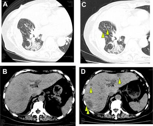
Figure S1 (A) Chest CT just before erlotinib monotherapy. (B) Abdominal CT just before erlotinib monotherapy revealed multiple liver metastases. (C) Chest CT at day 14 of erlotinib monotherapy. Pulmonary small nodules of right lower lobe appeared (arrowhead). (D) Abdominal CT at day 14 of erlotinib monotherapy revealed enlarged liver metastases (arrowhead).
Abbreviation: CT, computed tomography.
Disclosure
Drs Katsuyuki Kiura and Nagio Takigawa have received honoraria from Chugai Pharmaceutical and Sanofi-Aventis KK. The authors report no other conflicts of interest in this work.
Acknowledgments
The authors would like to thank Enago (http://www.enago.jp; Mumbai, India) for the English language review.