
Abstract
When Saccharomyces cerevisiae is starved of glucose, the gluconeogenic enzymes fructose-1,6-bisphosphatase (FBPase), malate dehydrogenase (MDH2), isocitrate lyase (Icl1) and phosphoenolpyruvate carboxykinase (Pck1) are induced. However, when glucose is added to prolonged starved cells, these enzymes are degraded in the vacuole via the vacuole import and degradation (Vid) pathway. Recent evidence suggests that the Vid pathway merges with the endocytic pathway at actin patches where endocytic vesicles are formed. The convergence of the Vid pathway with the endocytic pathway allows cells to remove intracellular and extracellular proteins simultaneously. However, the genes that regulate this step of the convergence have not been identified previously. Here we show that VID30 plays a critical role for the association of Vid vesicles and actin patches. Vid30 is constitutively expressed and interacts with Vid vesicle proteins Vid24 and Sec28 but not with the cargo protein FBPase. In the absence of SEC28 or VID24, Vid30 association with actin patches was prolonged. In cells lacking the VID30 gene, FBPase and Vid24 were not localized to actin patches, suggesting that Vid30 has a role in the association of Vid vesicles and actin patches. Vid30 contains a LisH and a CTLH domain, both of which are required for FBPase degradation. When these domains were deleted, FBPase trafficking to the vacuole was impaired. We suggest that Vid30 also has a role in the Vid pathway at a later step in a process that is mediated by the LisH and CTLH domains.
Acknowledgments
We thank Ryan Lucas for generating many of the reagents and plasmids used in this study. Primers were synthesized at the Core Facility of the Penn State University College of Medicine. This study was sponsored by an NIH grant R01 GM59480 and a Tobacco Settlement Fund to Hui-Ling Chiang.
Figures and Tables
Figure 1 VID30 is required for FBPase degradation in 3 d glucose starved cells. (A) Wild-type, vid30Δ, pre9D and blm10Δ mutant cells were starved for three d and transferred to medium containing fresh glucose for 0, 2 and 3 h. FBPase degradation was then determined. (B) Vid30 was tagged with HA and expressed in wild-type cells. Levels of FBPase and Vid30-HA in response to glucose re-feeding were determined. (C) Vid30 was tagged with V5-His and expressed in wild-type cells. FBPase degradation and Vid30-V5-His levels were examined. (D) Vid30-HA cells were glucose starved and shifted to glucose for 20 min. Levels of Vid30-HA, FBPase and Vid24-V5-His in the Vid vesicle enriched (V) and the cytosolic (C) fractions were examined. (E) Wild-type cells co-expressing Vid30-V5-His and Vid24-HA were glucose starved and shifted to glucose for 20 min. Vid30-V5-His was pulled down and the distribution of Vid30-V5-His, Vid24-HA, Sec28 and FBPase in the unbound (U) vs. bound (B) fractions was then determined. (F) Wild-type cells that co-expressed Vid24-V5-His and Vid30-HA were glucose starved and re-fed with glucose for 20 min and Vid24-V5-His was pulled down from total lysates. The distribution of Vid24-V5-His, Vid30-HA, Sec28 and FBPase in the unbound vs. bound fractions was examined. (G) Vid30-V5-His was co-expressed with Vid24-HA in the sec28D mutant that was starved for three d and then transferred to medium containing glucose for 20 min. Vid30-V5-His was pulled down and the presence of Vid24-HA and FBPase in the unbound vs. bound fractions was determined. (H) Vid30-V5-His was expressed in the vid24Δ mutant that was glucose starved and shifted to glucose for 20 min. Cells were lysed and Vid30-V5-His was pulled down. The presence of Vid30-V5-His, Sec28 and FBPase in the unbound and bound fractions was then determined. Relative ratios of protein levels were quantitated using ImageJ software.
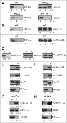
Figure 2 Abp1 and actin (Act1) are distributed in multiple locations. (A) Wild-type cells expressing Abp1-mCherry were glucose starved for 3 d. Cells were fractionated by differential centrifugation. The distribution of Act1 and Abp1-mCherry in the P1, P13, P100, P200 and S200 fractions was determined. (B) Abp1-mCherry was transformed into wild-type cells that expressed Sac6-GFP. The distribution of Abp1-mCherry and Sac6-GFP was determined. (C) Wild-type cells expressing Sac6-GFP was processed and actin patches were visualized with phalloidin conjugated with rhodamine. (D) Wild-type cells expressing Abp1-GFP was processed and actin patches were visualized with phalloidin conjugated with rhodamine.
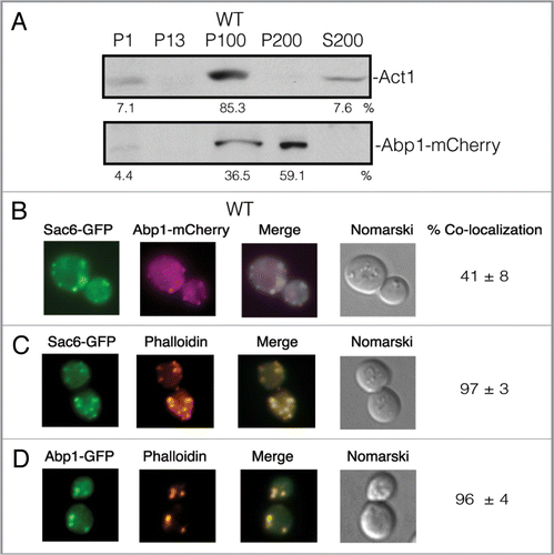
Figure 3 Vid30-GFP is associated with actin patches and FM-containing compartments. (A) Wild-type cells expressing Vid30-GFP were glucose starved and shifted to glucose for 0, 2 and 3 h. FBPase degradation was then examined. (B) The distribution of Vid30-GFP and actin patches was determined. (C) rvs161Δ cells expressing Vid30-GFP were re-fed with glucose and the distribution of Vid30-GFP and actin patches was examined. (D) Wild-type cells expressing Vid30-GFP were transferred to glucose-containing medium in the presence of FM for the indicated periods. The distribution of Vid30-GFP and FM was visualized using fluorescence microscopy.
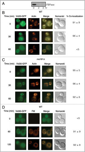
Figure 4 Vid30 association with actin patches is prolonged in the sec28Δ mutant and the vid24Δ mutant. (A) Vid30-GFP was expressed in the sec28Δ mutant that was glucose starved and then transferred to medium containing fresh glucose for the indicated time points. The distribution of Vid30-GFP with actin patches was determined. (B) Vid30-GFP was expressed in the vid24Δ mutant that was transferred from low to high glucose medium for the indicated time points. The distribution of Vid30-GFP with actin patches was examined.
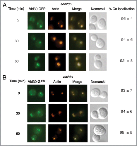
Figure 5 FBPase and Vid24 are present in the Vid vesicle enriched fraction in vid30Δ mutant cells. (A) Wild-type and the vid30Δ mutant cells were transferred to high glucose-containing medium for 20 min. The presence of FBPase in the Vid vesicle and cytosolic fractions was then determined. (B) Wild-type and vid30Δ cells expressing Vid24-HA were re-fed with glucose for 20 min. The distribution of Vid24 in the Vid vesicle and the cytosolic fractions was then examined. Protein levels were quantitated using ImageJ software.
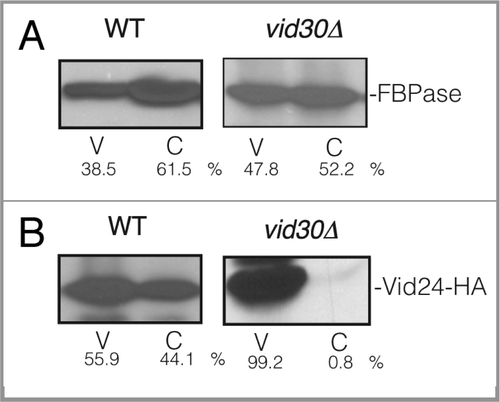
Figure 6 FBPase does not associate with actin patches in vid30Δ mutant cells. Wild-type (A) and vid30Δ mutant cells (B) expressing FBPase-GFP were glucose starved and re-fed with glucose for up to 60 min and examined by fluorescence microscopy for the distribution of FBPase-GFP and actin patches. (C) The vid30Δ mutant cells expressing FBPase-GFP were re-fed with glucose in the presence of FM for up to 120 min. The distribution of FBPase-GFP and FM was examined.
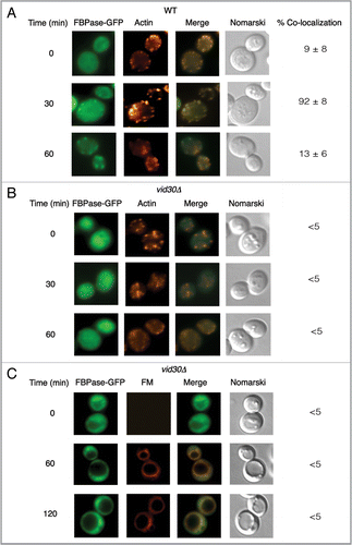
Figure 7 Vid24 does not associate with actin patches in cells lacking VID30. Wild-type (A) and vid30Δ mutant cells (B) expressing GFP-Vid24 were starved and replenished with glucose for up to 60 min and examined by fluorescence microscopy for the localization of GFP-Vid24 and actin patches. (C) The vid30Δ cell was re-fed with glucose and FM for up to 120 min and examined for the distribution of GFP-Vid24 and FM.
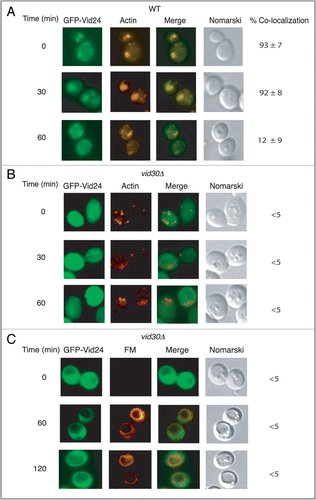
Figure 8 The LisH domain is required for FBPase degradation. (A) Schematic illustration of the position of the LisH and CTLH domains in Vid30 and the position of the LisH domain that was deleted in the ΔLisH mutant. (B) ΔLisH mutant cells were glucose starved and re-fed with glucose for up to 3 h. Levels of FBPase and ΔLisH-Vid30-V5-His were examined. (C) Cells co-expressing ΔLisH-Vid30-V5-His and Vid24-HA were starved and shifted to glucose for 20 min. ΔLisH-Vid30-V5-His was pulled down and the levels of Vid24-HA, Sec28 and FBPase in the bound vs. unbound fractions were then determined. (D) The ΔLisH mutant cells were shifted to glucose for 20 min. The distribution of ΔLisH-Vid30-V5-His, Vid24-HA and FBPase in the Vid vesicle and cytosolic fractions was examined. Relative ratios of proteins were quantitated using ImageJ software.
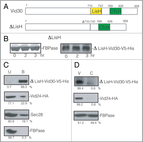
Figure 9 The ΔLisH mutant protein and FBPase accumulate in punctate structures in response to glucose. (A) ΔLisH-Vid30-GFP was expressed in yeast cells that were starved and re-fed with glucose for the indicated time points. The distribution of ΔLisH-Vid30-GFP and actin patches was determined by fluorescence microscopy. (B) The distribution of ΔLisH-Vid30-GFP and FM was examined. (C) FBPase-GFP was expressed in the ΔLisH mutant strain that was starved and re-fed with glucose. FBPase-GFP and FM was visualized by fluorescence microscopy.
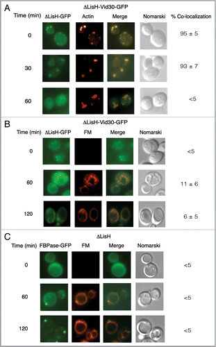
Figure 10 FBPase degradation is inhibited in the ΔCTLH mutant. (A) Schematic illustration of the position of the LisH and CTLH domains in Vid30 and the CTLH domain that was deleted to produce the CTLH domain mutant. (B) The CTLH domain of Vid30 was deleted and the mutant protein was tagged with V5-His. FBPase degradation was examined. The levels of ΔCTLH-Vid30-V5-His were determined. (C) Cells that co-expressed ΔCTLH-Vid30-V5-His and Vid24-HA were shifted to glucose for 20 min. Total lysates were prepared and ΔCTLH-Vid30-V5-His was pulled down. The presence of ΔCTLH-Vid30-V5-His, Vid24-HA, Sec28 and FBPase in the unbound and bound fractions was determined. (D) Cells that co-expressed ΔCTLH-Vid30-V5-His and Vid24-HA were glucose starved for three d and re-fed with glucose for 20 min. The distribution of ΔCTLH-Vid30-V5-His, Vid24-HA and FBPase in the Vid vesicle and cytosolic enriched fractions was examined and quantitated using ImageJ software.
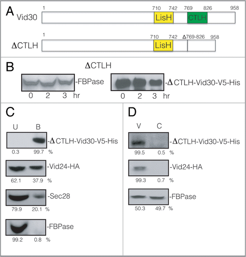
Figure 11 ΔCTLH-Vid30-GFP and FBPase accumulate in punctate compartments following glucose re-feeding. (A) ΔCTLH-Vid30-GFP was expressed in yeast cells that were starved and re-fed with glucose for the indicated times. ΔCTLH-Vid30-GFP and actin patches were visualized by fluorescence microscopy. (B) The distribution of ΔCTLH-Vid30-GFP and FM was examined. (C) FBPase-GFP was expressed in ΔCTLH mutant cells. The localization of FBPase-GFP and FM was then determined.
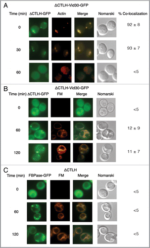
Figure 12 A model for VID30 in the Vid pathway. Vid vesicles associate with actin patches on the plasma membrane. Vid30 is present in Vid vesicles, actin patches, FM-containing endosomes and the vacuole. In the absence of Vid30, Vid vesicles fail to associate with actin patches. This suggests that Vid30 has a role in the association of Vid vesicles and actin patches. The absence of LisH or CTLH domain results in the distribution of these mutant proteins in punctate structures. When either the LisH or the CTLH domain is deleted, FBPase is localized in punctate structures. Because the patterns of FBPase distribution in the LisH and CTLH mutants are distinct from those seen in the complete absence of the VID30 gene, we suggest that these domains have a role in the Vid pathway at a later step.
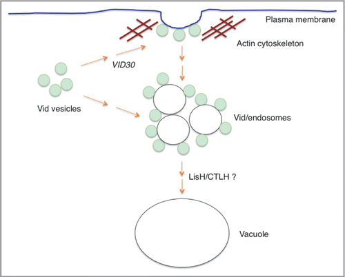
Table 1 Strains used in this study
Table 2 Primers used in this study