Abstract
Historically, ErbB3 has been overlooked within the ErbB receptor family due to its perceived lack of tyrosine kinase activity. We have previously demonstrated that in pancreatic cancer ErbB3 is the preferred dimerization partner of EGFR, ErbB3 protein expression level directly correlates with the anti-proliferative effect of erlotinib (an EGFR-specific tyrosine kinase inhibitor), and transient knockdown of ErbB3 expression results in acquired resistance to EGFR-targeted therapy. In this study, we develop a stable isogenic model of ErbB3 expression in an attempt to decipher ErbB3's true contribution to pancreatic cancer tumorigenesis and to examine how this receptor affects cellular sensitivity to EGFR-targeted therapy. Analysis of the EGFR-ErbB3 heterodimer demonstrates that ligand-induced PI3K-AKT signaling is limited to ErbB3-expressing cells and that this signaling cascade can be partially abrogated by inhibiting EGFR function with erlotinib. Using our model of exogenous ErbB3 expression we showed a direct relationship between ErbB3 protein levels and increased pancreatic cancer cell proliferation in vitro. In vivo, ErbB3+PANC-1 xenografts had a significantly larger tumor volume than PANC-1 control xenografts (ErbB3-PANC-1) and displayed increased sensitivity to EGFR-targeted therapy. In pancreatic cancer, ErbB3 appears to be critically involved in EGFR signaling as evidenced by its profound effect on cellular proliferation and its ability to influence response to EGFR-targeted therapy.
Acknowledgements
Financial support was received from a NIH/NRSA T32 Training Grant (J.S.L.) and from the Robert E. Reed Gastrointestinal Oncology Research Foundation.
Figures and Tables
Figure 1 Analysis of the transfected ErbB3 + PANC-1 cell line, confirms that ErbB3 is expressed, can dimerize, and can be phosphorylated by NRG-1β resulting in AKT signaling. (A) western blot analysis of ErbB3−PANC-1 and ErbB3+PANC-1 stable transfectant cells after stimulation with NRG-1β demonstrates expression and phosphorylation of ErbB3 in the ErbB3+PANC-1 cells. Significant increase in AKT activation is observed in the ErbB3+PANC-1 cell line. (B) western blotting for ErbB3 expression after covalently binding receptor dimers with Bis(Sulfosuccinimidyl)suberate (BS3) confirms the presence of ErbB3 dimerization within the ErbB3+PANC-1 cell line. As controls, ErbB3-associated dimers are absent in PANC-1 and present in AsPC-1 cells.
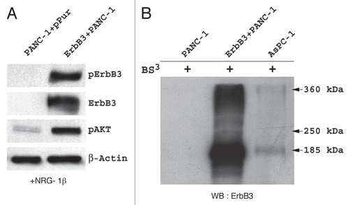
Figure 2 (A) Effect of ErbB3 expression on receptor heterodimerization and intra-cellular signaling in PANC-1 cells in the presence of EGF and NRG-1β stimulation. With ErbB3 present, EGF stimulation results in phosphorylation of EGFR and NRG-1β stimulation results in phosphorylation of ErbB3 as expected, but interestingly, in each scenario, activation of the non-target receptor is seen suggesting heterodimerization. Furthermore, ErbB3 expression permits AKT signaling with NRG-1β stimulation that, although diminished, persists in the presence of EGFR inhibition. ERK1/2 signaling is not effected by ErbB3 expression. (B) Western blotting of ErbB3 and EGFR expression in each of the 3 PANC-1 cell lines.
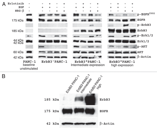
Figure 3 In PANC-1, increased ErbB3 expression directly correlates with increased cellular proliferation (p < 0.01) and sensitivity to EGFR targeted therapy (p < 0.01). (A) When allowed to propagate in 10% serum containing media, high-expressing ErbB3+PANC-1 cells and intermediate-expressing ErbB3+PANC-1 cells proliferated at rates 3 and 2.5 times that of ErbB3−PANC-1 cells, respectively, and these findings were observed after 3, 4 and 5 days of propagation. (B) The proliferative advantage of ErbB3 expression is also seen in serum-stressed conditions (0.5% FBS). (C) After 96 hours of erlotinib treatment, ErbB3+PANC-1 cells demonstrated significantly more growth inhibition relative to DMSO-treated controls than ErbB3-PANC-1 cells. Percent inhibition directly correlated with the level of ErbB3 expression (p < 0.05).
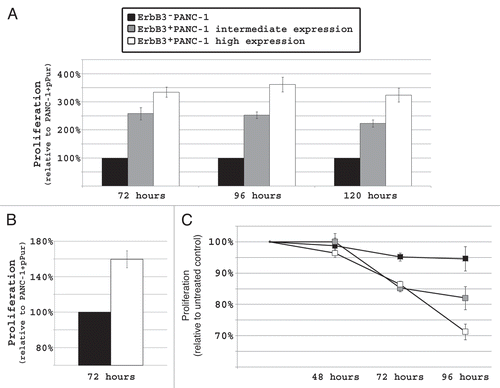
Figure 4 Inhibition of AKT signaling significantly diminishes PANC-1 cell proliferation. (A) western blot demonstrating that LY294002 (25 µmol/L) and PD98059 (15 µmol/L) successfully inhibits AKT and ERK1/2 signaling, respectively, in all 3 PANC-1 cell lines. (B) Dose effect of LY294002 and PD98059 on PANC-1 cell proliferation after 48 hours. LY294002 resulted in a significant decrease is proliferation (p < 0.05) relative to DMSO treated cells, while PD98059 has no inhibitory effect on proliferation of PANC-1 cells.
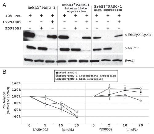
Figure 5 In PANC-1 xenografts, increased ErbB3 expression directly correlates with increased cellular proliferation (p < 0.05) and sensitivity to EGFR targeted therapy (p < 0.05). (A) After 5 weeks, ErbB3+PANC-1 xenografts had a significantly larger mean tumor volume (479.6 ± 60.7 mm3 vs. 261.1 ± 35.0 mm3; p < 0.05). (B) When treated with erlotinib, ErbB3+PANC-1 xenografts demonstrated a significant greater decrease in the rate of proliferation than did ErbB3−PANC-1 xenografts relative to vehicle-treated control groups. Tumor growth in each cell line is plotted with vehicle treated controls to demonstrate that ErbB3+PANC-1 xenografts displayed increased tumor proliferation, and that when treated with erlotinib, ErbB3+PANC-1 xenografts were not significantly larger than ErbB3−PANC-1 treated tumors. Data represents the mean ± SEM for 8 xenografts. (C) Western blot xenograft analysis confirms ErbB3 expression and shows that erlotinib results in diminished phospho-ErbB3 and phospho-AKT signaling in ErbB3+PANC-1 tumors.
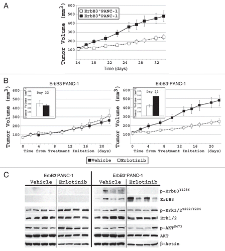
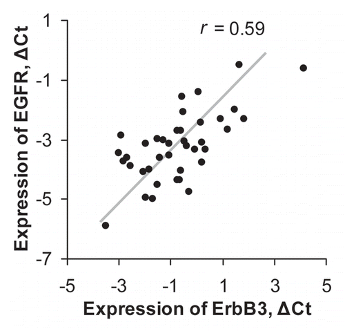
Figure 6 Expression levels of ErbB3 and EGFR mRNA in pancreatic cancer surgical specimens for 39 patients. ΔCt is calculated relative to expression of RPLPO.r is a Spearman correlation coefficient.