
Abstract
Comment on: Gavathiotis E, et al. Mol Cell 2010; 40:481-92.
Figures and Tables
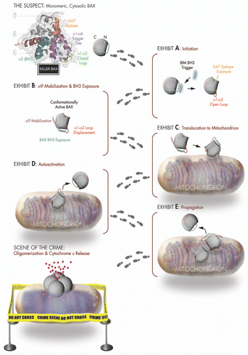
Figure 1 BH3-triggered structural reorganization drives the activation of pro-apoptotic BAX. Killer BAX is disguised as an inactive cytosolic protein (see mug shot of “The Suspect”) and is coerced to undergo a series of conformational changes upon engagement of its α1/α6 binding site (purple) by a triggering BH3 α-helix. BAX's transformation from an innocent to a guilty protein begins with displacement of its α1–α2 loop from a closed (green) to an open (red) position (Exhibit A), which reveals the 6A7 epitope (orange) and leads to mobilization of the C-terminal α9 helix (maroon) for mitochondrial translocation and exposure of the BAX BH3 death domain (cyan) (Exhibits B and C). BAX propagates its own activation through triggering interactions between the exposed BAX BH3 α-helix of fully activated monomers and the α1/α6 binding site of inactive monomers (Exhibits D and E), ultimately assembling into an elusive oligomeric pore that promotes apoptosis by releasing mitochondrial accomplices such as cytochrome c at the “Scene of the Crime.”
Comment on: Gavathiotis E, et al. Mol Cell 2010; 40:481 - 492