Abstract
The development of bilateral symmetry during the evolution of species probably 600 million years ago brought about several important innovations: It fostered efficient locomotion, streamlining, and favored the development of a central nervous system through cephalization. However, to increase their functional capacities, many organisms exhibit chirality by breaking their superficial left-right (l-r) symmetry, which manifests in the lateralization of the nervous system or the l-r asymmetry of internal organs. In most bilateria, the mechanisms that maintain consistent l-r asymmetry throughout development are poorly understood. This review highlights insights into mechanisms that couple early embryonic l-r symmetry breaking to subsequent l-r patterning in the roundworm Caenorhabditis elegans. A recently identified strategy for l-r patterning in the early C. elegans embryo is discussed, the spatial separation of midline and anteroposterior axis, which relies on a rotational cellular rearrangement and non-canonical Wnt signaling. Evidence for a general relevance of rotational/torsional rearrangements during organismal l-r patterning and for non-canonical Wnt signaling/planar cell polarity as a common signaling mechanism to maintain l-r asymmetry is presented.
Acknowledgements
The author would like to thank Zhirong Bao for continuous support and Antony Santella for critical reading of the manuscript. The author is supported by a long-term fellowship from the Human Frontier in Science Program Organization.
Figures and Tables
Figure 1 L-R Asymmetry of internal organs in Caenorhabditis elegans, Drosophila melanogaster and Homo sapiens. Selected organs are shown for each organism, endodermal/intestinal organs are shown in red. Lower right of each part: Schematized topology of selected asymmetries for each organism. Arrows indicate tissue movements that lead to coiling of the respective tissue.

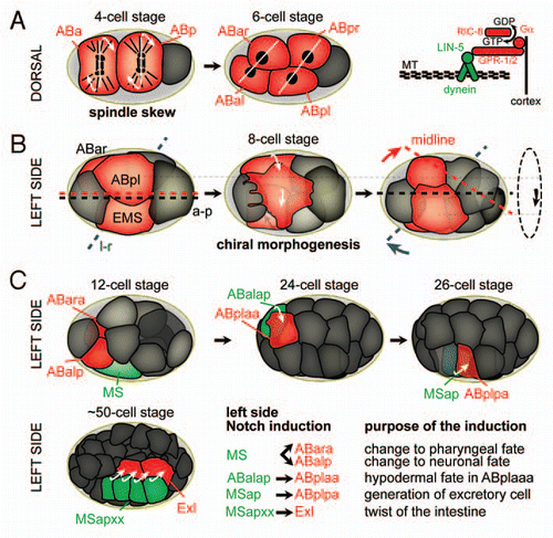
Figure 2 The sequence of l-r symmetry breaking events during C. elegans embryogenesis. Cells actively involved in morphogenetic events or inductions are highlighted in red. (A) Left: Spindle skew during the ABa/p divisions that leads to the first morphological l-r asymmetry. Dorsal views, anterior is to the left, posterior to the right. White lines connect daughter nuclei, arrows indicate the direction of spindle skewing. Right: The force-generating machinery that links the spindle to the cortex. Gα (GPA-16 and GOA-1, see text for details) recruit the dynein complex, a minus-end directed microtubule motor, through interactions with GPR-1/2 and the coiled-coil protein LIN-5. MT, microtubules. (B) Chiral morphogenesis at the 8-cell stage. Left side views, anterior is to the left. Midline (red), a-p axis (black) and l-r axis (blue) are shown. The dashed circle on the right indicates the direction of the rotational rearrangement. (c) Notch inductions that follow chiral morphogenesis on the left side of the embryo. Embryos are oriented as in b. Inducing cells are shown in green, receiving cells in red. For simplicity, lateral cells were obmitted in the case of the Notch induction from MSapxx, as this induction takes place in the center of the embryo.