
Abstract
Ch14.18 is a mouse-human chimeric monoclonal antibody to the disialoganglioside (GD2) glycolipid. In the clinic, this antibody has been shown to be effective in the treatment of children with high-risk neuroblastoma, either alone or in combination therapy. Extensive product characterization is a prerequisite to addressing the potential issues of product variability associated with process changes and manufacturing scale-up. Charge heterogeneity, glycosylation profile, molecular state and aggregation, interaction (affinity) with Fcγ receptors and functional or biological activities are a few of the critical characterization assays for assessing product comparability for this antibody. In this article, we describe the in-house development and qualification of imaged capillary isoelectric focusing to assess charge heterogeneity, analytical size exclusion chromatography with online static and dynamic light scattering (DLS), batch mode DLS for aggregate detection, biosensor (surface plasmon resonance)-based Fcγ receptor antibody interaction kinetics, N-glycoprofiling with PNGase F digestion, 2-aminobenzoic acid labeling and high performance liquid chromatography and N-glycan analysis using capillary electrophoresis. In addition, we studied selected biological activity assays, such as complement-dependent cytotoxicity. The consistency and reproducibility of the assays are established by comparing the intra-day and inter-day assay results. Applications of the methodologies to address stability or changes in product characteristics are also reported. The study results reveal that the ch14.18 clinical product formulated in phosphate-buffered saline at a concentration of 5 mg/ml and stored at 2–8°C is stable for more than five years.
Acknowledgments
This project has been funded in whole or in part with federal funds from the National Cancer Institute, National Institutes of Health, under Contract No. HHSN261200800001E. The content of this publication does not necessarily reflect the views or policies of the Department of Health and Human Services, nor does mention of trade names, commercial products or organizations imply endorsement by the US government. This research was supported in part by the Developmental Therapeutics Program in the Division of Cancer Treatment and Diagnosis of the National Cancer Institute.
We would like to acknowledge the support and encouragement from Dr. Stephen P. Creekmore (chief of Biological Resources Branch of NCI) and Dr. Karen Muszynski (Program Director, Biological Resources Branch, NCI). We would also like to acknowledge the technical support from Ms. Eying Chen during the initial stages of this work. The generous supply of purified clinical grade ch14.18 from the Biopharmaceutical Development Program (BDP; SAIC-Frederick, Inc.,) manufacturing group is greatly appreciated. We also would like to thank the BDP process analytics group for providing the infusion mock model processed samples.
Figures and Tables
Figure 1 Imaged cIEF profiling of ch14.18. Imaged cIEF profiling was performed as described in Materials and Methods. Four different lots of ch14.18 (labeled L1, L2, L3 and L4) manufactured at different times using the same manufacturing procedure were analyzed. Overlay of the profiles is shown.
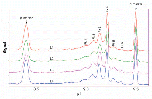
Figure 2 Analytical SEC-MALS profile of ch14.18. SEC-MALS and data analysis are performed as described in Materials and Methods. (A) is light-scattering signal overlay and (B) is UV280 signal overlay. (C) is batch mode DLS of ch14.18. Shown is a typical DLS overlay of the % mass vs. radius plot of ch14.18. DLS of eight sample vials (reference standard, samples control vials retained under stability control in-house and sample material retained after use in the clinic and returned for test). The overlay includes samples scanned directly and after filtration through 0.2 µm filtration.
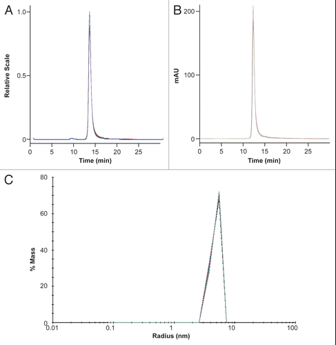
Figure 3 N-glycosyl profile of ch14.18. N-glycoprofiling was performed following the procedure described in Materials and Methods. (A) Overlay of the HPLC profile of the same lot from independent analysis. (B) Overlay of HPLC profile of two separate lots of ch14.18.
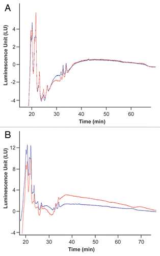
Figure 4 N-glycan analysis of ch14.18. (A) Comparison of N-glycan profile ch14.18, control human IgG1(κ) and murine IgG1(κ). Experimental details are described in Materials and Methods. (B) Intra-day and inter-day consistency in the N-glycan profile of ch14.18. Data were collected from analysis of a reference lot ch14.18 from different experiments performed within the same day or on different days and plotted. The mean of all the estimates and the standard deviation from average are also indicated.
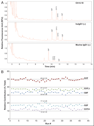
Figure 5 Sensorgrams showing dose-dependent binding of ch14.18 (A) and a control human IgG1(κ) (B) on FcγRIIIA coupled to a CM-5 chip. FcγRIIIA binding kinetics was performed as described in Materials and Methods. Serial 2-fold dilutions (eight) of ch14.18 or control IgG1(κ), starting from 5 µM (curve 1) to 39.8 nM (curve 8), were injected.
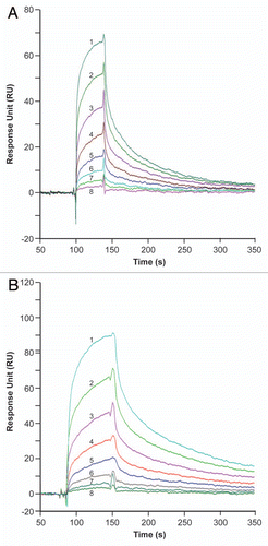
Figure 6 Ch14.18-mediated CDC determined by MTS colorimetric cell proliferation inhibition assay. (A) Cell specificity; (B) ch14.18 specificity. Assay was performed as described in Materials and Methods.
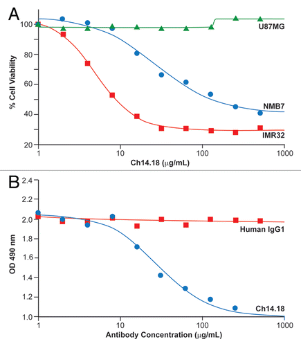
Figure 7 Binding specificity of ch14.18 probed by CbELISA. (A) CbELISA specificity of ch14.18 binding to GD2 positive cells. CbELISA was performed as described previously in reference Citation28. (B) GD2 specificity of FITC-labeled ch14.18 binding cellular specificity determined by cell sorting using FACS analysis. Cells were stained with FITC labeled ch14.18 (6 µg/ml) and FACS analysis performed as described in Materials and Methods. (C) FITC-ch14.18 dose-dependent shift in FACS sorting of GD2-expressing IMR32 neuronal cell. Assays were performed as described in Materials and Methods. Control has no ch14.18 added. Histograms 1, 2, 3 and 4 have 1, 3, 9 and 10 µg FITC-labeled ch14.18, respectively.
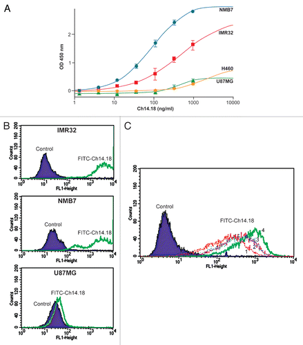
Figure 8 (A) Effect of acid (low pH) and high temperature on the molecular state of ch14.18. SEC-HPLC was performed as described in Materials and Methods. (B) Change in molecular state of ch14.18 on high temperature treatment parallels the CDC effect. (C) CDC effect of ch14.18 is demonstrated using cell death probed by FACS analysis using propidium iodide. Experimental details are described in Materials and Methods.
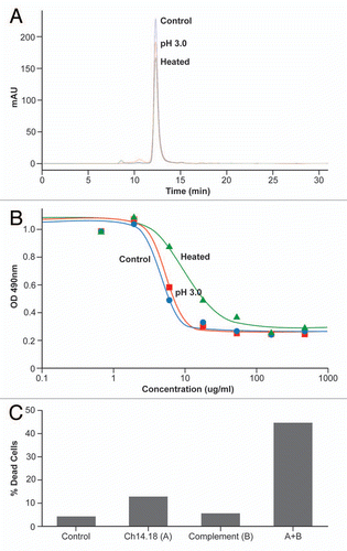
Table 1 pI and relative % peak area for different clinical lots of ch14.18
Table 2 Inter-day variations in the relative peak areas of the components of ch14.18 reference standard lot
Table 3 Relative peak area % of total area for each integrated peak of all the post-administration clinical vial samples shipped back from clinical sites
Table 4A Inter-day variation in the molecular weight estimates of albumin standard
Table 4B Estimated molecular parameters of ch14.18 reference standard from SEC-MALS experiments
Table 4C Molecular state of ch14.18 control and infusion model mock samples during 24 h mock infusion model study
Table 5 Intra-day and inter-day variations in the equilibrium dissociation constant of the FcγRIIIA receptor binding interaction of ch14.18
Table 6A Intra-day and inter-day variations in the ch14.18-mediated CDC of NMB7 cells with human and rabbit complement sera
Table 6B Relative CDC of two different lots of ch14.18 compared with the CDC of a reference lot of ch14.18
Table 7 Effect of acid and heat treatment on the SEC profile of ch14.18