
Abstract
For directional cell migration to occur cells must interpret guiding cues present in their environment. Chemotaxis based on negative or positive signals has been long thought as the main driving force of guided cell migration. However during collective cell migration cells do receive information from external signals but also upon interactions with their direct neighbours. These multiple inputs must be translated into intracellular reorganisation in order to promote efficient directional migration. Small GTPases, being involved in establishing cell polarity and regulating protrusive activity, are likely to play a central role in signal integration. Indeed, recent findings from our laboratory indicate that Contact-Inhibition of Locomotion controlled by N-Cadherin and chemotaxis dependent on Sdf1/Cxcr4 signalling converge towards regulation of the localized activity of RhoA and Rac1. All together they establish cell polarity and select well-oriented cell protrusions to ensure directional cell migration.
Figures and Tables
Figure 1 Signal integration through RhoGTPases during collective chemotaxis in neural crest cells. (A) Representation of the border of a neural crest cell group. Cells are polarized according to their cell contacts by a mechanism dependent on CIL.Citation8,Citation10 RhoA and Rac1 activity are shown in red and green respectively. Only outer cells have a free edge and exhibit a clear front-back polarity. Inner cells remain unpolarized. (B) Intracellular signaling integrating inputs from contact-inhibition of locomotion and chemotaxis through small GTPases. At the cell contact N-cadherin/Syndecan-4/PCP (Frizzled-Dsh)/CIL signaling leads to a strong RhoA activity restricting Rac1 at the free-edge. At the back, RhoA/ROCK signaling controls stress fibers formation and cell body contraction in part through regulation of myosin light chain (MLC) phosphorylation. At the front, Rac1 activity controls WAVE/Arp2/3 cascade leading to actin branching and lamellipodium formation/stabilization and antagonizes RhoA impact on MLC via activation of PAK. In addition, Rac1 activity may be amplified by a PI3K/GEFs/Rac1 positive feedback loop downstream of Cxcr4. (C) Summary of the main players involved in establishing and maintaining front and back cell identities. Arp2/3, Actin-related proteins 2/3; Dsh, Dishevelled; PAK, p21-activated kinases; ROCK, Rho-Associated Kinase; WAVE, WASP family verprolin-homologous. Other abbreviations have been described in the text.
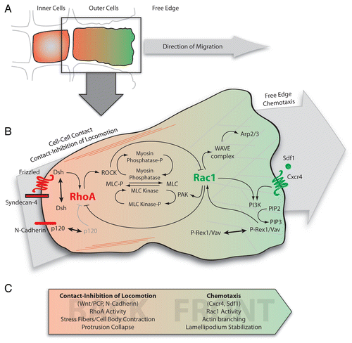
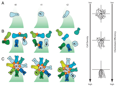
Figure 2 Different migratory behaviours of neural crest cells placed in a gradient of Sdf1. (A) Single cells that are isolated from other neural crest cells fail to polarize according to the Sdf1 gradient and therefore show poor chemotaxis. (B) Single cells that experience transient contacts are polarized upon collisions and chemotax more or less efficiently according to cell density. (C) Cell clusters show a radial symmetry with cells polarized along their cell contact—free edge axis. Front cell protrusions are stabilized and generate a driving force towards Sdf1. Orientation and size of the arrows indicate the direction and stability of cell protrusions. Round arrows mark tumbling and non-polarized cells. Cells are colour coded to help follow their behaviour from one time point to the other. (D) Typical cell tracks obtained in each situation showing that chemotaxis improves as cell density increases. Cell collisions are shown as stars. Shades of green represent Sdf1 gradient. Based on data from reference Citation6.
Extra View to: Theveneau E, Marchant L, Kuriyama S, Gull M, Moepps B, Parsons M, Mayor R. Collective chemotaxis requires contact-dependent cell polarity. Dev Cell 2010; 19:39 - 53; PMID: 20643349; http://dx.doi.org/10.1016/j.devcel.2010.06.012