
Abstract
Vibrioparahaemolyticus ExsA is the transcriptional regulator for type III secretion system 1 (T3SS1) while ExsD blocks T3SS1 expression. Herein we show that deletion of exsC from V. parahaemolyticus blocked synthesis of T3SS1-dependent proteins under inducing conditions (contact with HeLa cells), while in trans complementation of the ΔexsC strain with wild-type exsC restored protein synthesis. Under non-inducing conditions (Luria broth plus salt), in trans expression of exsC in a wild-type strain resulted in synthesis and secretion of T3SS1-dependent proteins. Deletion of exsC does not affect the synthesis of ExsA while expression of T3SS1 genes is independent of ExsC in the absence of ExsD. Co-expression of recombinant proteins with different antigenic tags demonstrated that ExsC binds ExsD and that the N-terminal amino acids of ExsC (positions 7 to 12) are required for binding. Co-expression and purification of antigentically tagged ExsA and ExsD demonstrated that ExsD directly binds ExsA and presumably prevents ExsA from binding promoter regions of T3SS1 genes. Collectively these data demonstrate that ExsD binds ExsA to block expression of T3SS1 genes, while ExsC binds ExsD to permit expression of T3SS1 genes. ExsA, ExsC, and ExsD from V. parahaemolyticus appear to be functional orthologues of their Pseudomonasaeruginosa counterparts.
Acknowledgements
We gratefully acknowledge Lisa Orfe, Patrick Friel, Daniel Erwin, Seth Nydam and Pablo Piñeyro for their technical assistance and discussions. Dr. Kathryn J. Boor provided the wild-type strain of V. parahaemolyticus (NY-4). This project was supported in part by National Institute of Health, Department of Health and Human Services under the contract number NO1-AI-30055 and by the Agricultural Animal Health Program, College of Veterinary Medicine, Washington State University.
Figures and Tables
Figure 1 ExsC is required for the synthesis of Vp1656 and Vp1659 upon V. parahaemolyticus infection of HeLa or Caco-2 cells. Wild-type (NY-4) (lanes 1 and 3) and ΔexsC (lanes 2 and 4) strains were used to infect HeLa (Lanes 1 and 2) and Caco-2 (lanes 3 and 4) cells for 4 h. The whole-cell lysates of the infected samples were probed with polyclonal antibody against Vp1656 (upper), Vp1659 (middle) and for the DNA loading control DnaK (lower).
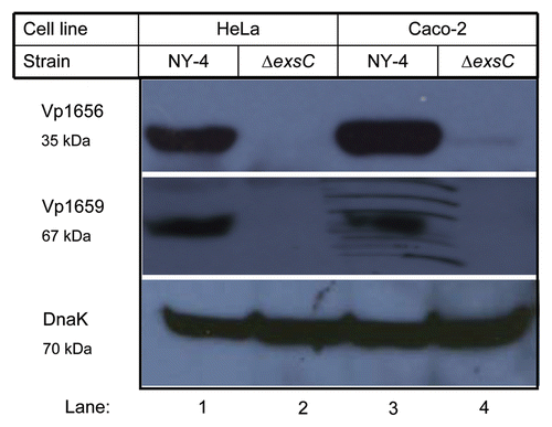
Figure 2 Western blot of whole-cell lysates showing that in trans expression of exsC activates production of Vp1656 and Vp1659. The NY-4 and ΔexsC strains were transformed with an ExsC expression plasmid or control plasmid (pMMB208-bla) and were then grown in non-inducing (LB-S) or inducing conditions (HeLa cell infection) (4 hr). Analysis of the whole-cell lysate showed that NY-4 did not synthesize Vp1656 or Vp1659 when grown in LB (Lane 1) unless ExsC was expressed in trans (Lane 3). In trans expression of exsC led to higher production of both Vp1656 and Vp1659 (Lane 4) compared to wild-type expression levels (Lane 2). A similar pattern of expression (Lanes 5–8) was evident with in trans expression of ExsC for the ΔexsC strain.
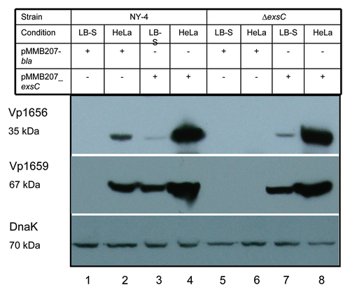
Figure 3 Analysis of secreted proteins shows that ExsC activates secretion of Vp1656 and Vp1659 when grown in non-inducing conditions (LB-S). Proteins were precipitated from the supernatant of NY-4 (lane 1), NY-4:pMMB207_exsC (lane 2), NY-4:pMMB207_bla (lane 3), NY-4:pexsA (lane 4) and ΔexsD (lane 5) strains and probed with polyclonal antibody against Vp1656 (upper), Vp1659 (middle) and DnaK (lower).
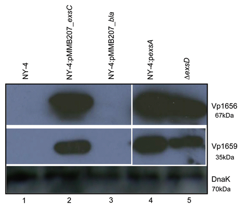
Figure 4 Deletion of ExsC does not reduce the synthesis of ExsA under inducing conditions. For these experiments exsA (chromosomal) was tagged with the HA antigen sequence in both the NY-4 (lanes 1 and 2) and ΔexsC (lanes 3 and 4) strains. After growth in LB-S (lanes 1 and 3) for 4 h or with HeLa cells (lanes 2 and 4) for 4 h, whole-cell lysates were collected and probed with anti-HA (upper) or anti-DnaK (lower) by western blot analysis.
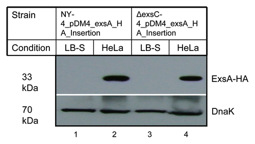
Figure 5 ExsC is not required for the expression of T3SS1 genes in the ΔexsD strain. (A) Protein samples isolated from ΔexsD exsC (lane 1) and ΔexsD (lane 2) strains grown in LB-S were probed with polyclonal antibody against Vp1656 (upper), Vp1659 (middle) and DnaK (lower); (B) RT-PCR analysis was used to detect mRNA transcripts from a subset of T3SS1 genes for the ΔexsD exsC (lane 1) and ΔexsD strains grown in LB-S.
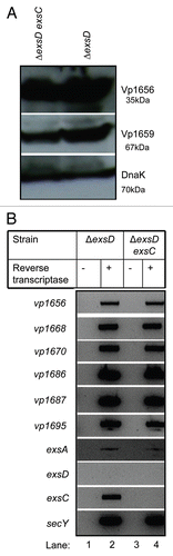
Figure 6 ExsC binds ExsD when these proteins are expressed concurrently. Whole-cell lysates from NY-4:pexsD-HA-pexsC-His (lane 1), NY-4:pexsD-HA (lane 4) were probed with anti-HA (upper; ExsD) and anti-His (lower; ExsC). After passage through a nickel column (which binds the His tag) and washing, eluted fractions from NY-4:pexsD-HA-pexsC-His (lanes 2 and 3), NY-4:pexsD-HA (lanes 5 and 6) were probed with anti-HA (upper) and anti-His (lower) antibodies. ExsC and ExsD were eluted together (lanes 2 and 3) and this outcome was not the result of non-specific binding of the HA protein and the nickel column (lanes 5 and 6).
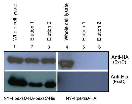
Figure 7 The N-terminus of ExsC is required for ExsC to bind with ExsD. (A) Schematic representation of fusion proteins between full-length exsC and GST (upper), and between N-terminus truncated exsC and GST (lower). “Deletion” indicates the location of the 5-prime deletion for the truncated exsC sequence. (B) Whole-cell lysate and elution from NY-4:pGST-exsC-His pexsD-HA or from NY-4:pGST_exsC_Δ5′-His pexsD-HA strains probed with anti-His (upper) or anti-HA (lower) antibodies. The truncated ExsC_Δ5′protein does not bind ExsD.
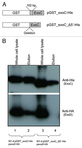
Figure 8 Full-length ExsC is required for transcription and expression of T3SS1 genes. (A) Strains were cultured separately in LB-S and then cell lysates from NY-4:pGST_exsC-_Δ5′-His (lane 1) and NY-4:pGST-exsC-His (lane 2) were probed with anti-Vp1656, anti-Vp1659, anti-His and anti-DnaK; (B) RT-PCR analysis showing mRNA transcripts for a panel of T3SS1 genes for the NY-4:pGST-exsC-His and NY-4:pGST_exsC_Δ5′-His strains after growth in LB-S. SecY, a housekeeping gene, served as a positive control for the RT-PCR experiment.
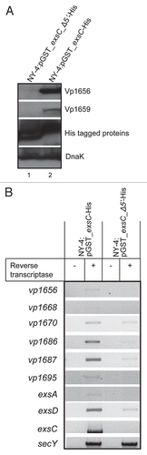
Figure 9 Amino acids from position 7 to 12 of ExsC are required for ExsC to interact with ExsD. Whole-cell lysate and eluted fractions from NY-4:pGST-exsC(7-148)-His pexsD-HA, NY-4:pGST-exsC(13-148)-His pexsD-HA or NY-4:pGST-exsC (27-148)-His pexsD-HA strains were probed with anti-HA (lower) and anti-His (upper) antibodies. “W” indicates whole-cell lysates and “E” indicates eluted fraction.
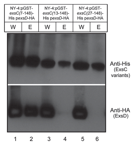
Figure 10 ExsD binds ExsA when expressed concurrently in vivo while GST-fused ExsC does not bind ExsA. Western blots of NY-4 grown in LB-S with co-expression of ExsA with either (A) GST_ExsC_6xHis, or (B) ExsD_HA. ExsA_HA was not eluted with GST_ExsC_6xHis, but ExsD_HA was eluted with ExsA-6xHis indicating that ExsC does not bind ExsA while ExsD does bind ExsA. Specificity of the nickel column for isolation of 6xHis tagged proteins is shown in .
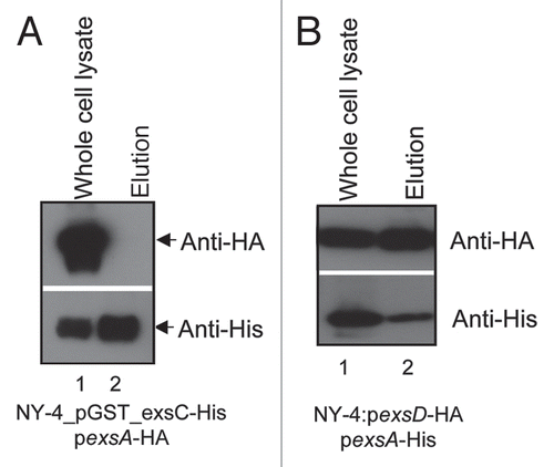
Figure 11 A model illustrating the regulation of T3SS1 genes. When ExsD is present in molar excess of ExsC, ExsD binds ExsA and blocks transcriptional promoter activity. T3SS1 genes are expressed when ExsC binds ExsD or if ExsA is synthesized to a molar concentration in excess of the ExsD concentration. The induction signal for ExsC has not been identified and there is no direct evidence whether or not the same or independent induction signal is required to express and synthesize ExsA (the latter can occur independently of ExsC; ).
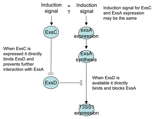
Table 1 Strains and plasmids used in this study
Table 2 Primers used in this study