
Abstract
Candida albicans can develop biofilms on medical devices and these biofilms are most often nourished by a continuous flow of body fluids and subjected to shear stress forces. While many C. albicans biofilm studies have been carried out using in vitro static models, more limited information is available for biofilms developed under conditions of flow. We have previously described a simple flow biofilm model (SFB) for the development of C. albicans biofilms under conditions of continuous media flow. Here, we recount in detail from a methodological perspective, this model that can be assembled easily using materials commonly available in most microbiological laboratories.The entire procedure takes approximately two days to complete. Biofilms developed using this system are robust, and particularly suitable for studies requiring large amounts of biofilm cells for downstream analyses. This methodology simplifies biofilm formation under continuous replenishment of nutrients. Moreover, this technique mimics in vivo flow conditions, thereby making it physiologically more relevant than the currently dominant static models.
Acknowledgements
Biofilm-related work in the laboratory is funded by grants numbered R21DE017294 and R21AI080930 from the National Institute of Dental & Craniofacial Research and the National Institute for Allergy and Infectious Diseases (to J.L.L.-R.). P.U. is supported by a postdoctoral fellowship, 10POST4280033, from the American Heart Association. The content is solely the responsibility of the authors and does not necessarily represent the official views of the NIDCR, the NIAID, the NIH or the AHA.
Figures and Tables
Figure 1 (A) Picture of the fully assembled apparatus inside the incubator. (B) A close-up picture of the tube with the cut-off bottom and the inserted tubing.
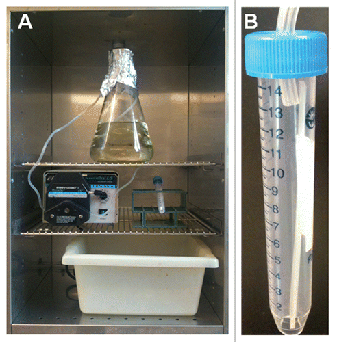
Figure 2 Macroscopic and microscopic observations of biofilms grown in the simple flow model. (A) Macroscopically, biofilms grown in the flow biofilm model appeared mucoid and highly wrinkled in appearance. (B) Confocal laser scanning microscopy reveals a thick biofilm with a highly filamentous top layer.

Table 1 Potential problems and advice on possible solutions
Table 2 Results of antifungal susceptibility testing of biofilms formed under conditions of flow by C. albicans type strain SC5314 against fluconazole, amphotericin B and caspofungin