
Abstract
Transmissible Spongiform Encephalopathy (TSE) agents are defined by their virulence for particular species, their spread in the population, their incubation time to cause disease, and their neuropathological sequelae. Murine adapted human agents, including sporadic CJD (sCJD), New Guinea kuru, and Japanese CJD agents, display particularly distinct incubation times and maximal infectious brain titers. They also induce agent-specific patterns of neurodegeneration. When these TSE agents are transmitted to cultured hypothalamic GT1 cells they maintain their unique identities. Nevertheless, the human kuru (kCJD) and Japanese FU-CJD agents, as well as the sheep 22L and 263K scrapie agents display doubling times that are 8x to 33x faster in cells than in brain, indicating release from complex innate immune responses. These data are most consistent with a foreign viral structure, rather than an infectious form of host prion protein (PrP-res). Profound agent-specific inhibitory effects are also apparent in GT1 cells, and maximal titer plateau in kCJD and FU-CJD differed by 1,000-fold in a cell-based assay. Remarkably, the lower titer kCJD agent rapidly induced de novo PrP-res in GT1 cells, whereas the high titer FU-CJD agent replicated silently for multiple passages. Although PrP-res is often considered to be toxic, PrP-res instead may be part of a primal defense and/or clearance mechanism against TSE environmental agents. Limited spread of particular TSE agents through nanotubes and cell-to-cell contacts probably underlies the long peripheral phase of human CJD.
Acknowledgments
This work was supported by NIH grants NINDS ROI 012674 and NIAID grant R21 A1076645.
Figures and Tables
Figure 1 Infectivity assays of FU-infected brain homogenates. (A) Western blots of GT1 indicator cells exposed to serial FU-CJD brain dilutions (3e5 to 3 CE) at p5; with dilution decreasing amounts of PrP-res are induced. Proteinase K treatment (PK) and relative protein load indicated in each lane are indicated, as well as the %PrP-res (of total PrP) beneath the +PK lanes. Note the FU-CJD diagnostic 13 kDa PrP-res band (arrow). (B) With further passages, PrP-res signals are detectable with an input of only 3CE as shown at p13. Since additional passages did not show a PrP-res signal with lower input CE, FU-CJD homogenates have 1 TCID per 3 brain cells. (C). Graph of %PrP-res induced by each serial CE dilution at progressive passages. Note that the incubation time curves are roughly parallel for each dilution, i.e., the PrP-res rate of increase is similar for each input dilution. PrP-res in FU-CJD cells became maximal at ∼25%, and minor PrP-res variations between 25–31% were due to variable breakdown during cell collections; when cellular lysosomal enzymes digest PrP during collection, total PrP will be reduced, and thus the %PrP-res falsely increased. (D) Plots the log agent TCID to the linear accumulation of %PrP-res at different passages. More extensive passages with 3 CE input (p15) confirmed a 4 log discrimination of the FU-CJD agent by this assay. This as well as other western blots shown here revealed no additional small bands of <12 kd as previously depicted in references Citation3 and Citation4.
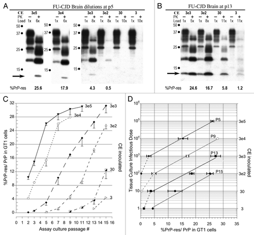
Figure 2 Infectivity of GT1 cells chronically infected with FU-CJD. (A) Representative western blots of GT1 homogenate dilutions at p7 and p13. Progressive passages show infectivity in as little as 0.3 CE by p13. This is a 10 fold higher TCID than FU-CJD brain (). PK treatment, CE applied, %PrP-res induced with each serial dilution, and the diagnostic 13 kDa band are indicated. Note the GT1 cells exposed to FU-infected cell homogenate dilutions accumulate PrP-res more rapidly than those exposed to FU-infected brain homogenates. (B) Graph of progressive PrP-res accumulation at each input CE dilution. (C) Standard plots of log TCID to %PrP-res shows the same slope in FU-CJD infected cells and brain; the only difference is that fewer infected cells were required to induce the same %PrP-res, i.e., the cells were consistently more infectious.
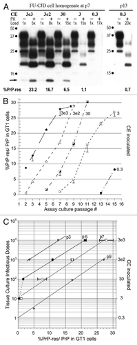
Figure 3 Western blot TCID assays of kCJD-infected brain. (A) Passage number, input cell equivalents (CE) and relative protein loads applied to each lane are indicated. The reference brain-specific PrP-res band pattern loaded with 3e5 brain homogenate CE is not visible in the GT1 indicator cells exposed to kCJD brain. Instead, a de novo GT1-specific PrP-res pattern is already seen at p1. A series of input dilutions, including lower CE are shown for p7 and p13 after kCJD brain infection. Note at p1 and p7 the %PrP-res is the same and does not rise, e.g., compare 3e4 and 3e3 CE inputs at p1 and p7. The %PrP-res in GT1 cells exposed to 300 CE was detectable on only very dark films at p7 but was clearly positive at p13 with a 17× load. Thus kCJD brain contained maximally 1 TCID per 300 cells, a 1,000-fold lower titer than FU-CJD-infected cells. The mouse brain PrP-res pattern was the same for kCJD and FU-CJD as shown previously in references Citation3 and Citation4 and in those transmissions, no bands of <15 kd were seen in kCJD infected GT1 cells. (B) Plot of %PrP-res exposed to CE dilutions at sequential passages confirms the lack of increase in %PrP-res with time. Nevertheless, the different levels of %PrP-res accurately discriminate different input CE s (and titer). There is a significant difference in the %PrP-res from 300 to 3e5 CE at p7, yielding a 3 log TCID span for kCJD. (C) Standard kCJD plot for %PrP-res to infectious titer. Note that only the same slope, but the same line is seen with different inputs at p5 and p7.
Table 1 TSE agents display very divergent replication “clocks” when studied in the same host background as seen in the above numbers