Abstract
Many bacterial species use secreted quorum-sensing autoinducer molecules to regulate cell density- and growth phase-dependent gene expression, including virulence factor production, as sufficient environmental autoinducer concentrations are achieved. Bacillus anthracis, the causative agent of anthrax, contains a functional autoinducer (AI-2) system, which appears to regulate virulence gene expression. To determine if the AI-2 system is necessary for disease, we constructed a LuxS AI-2 synthase-deficient mutant in the virulent Ames strain of B. anthracis. We found that growth of the LuxS-deficient mutant was inhibited and sporulation was delayed when compared with the parental strain. However, spores of the Ames luxS mutant remained fully virulent in both mice and guinea pigs.
Introduction
Bacillus anthracis is a rod-shaped, Gram-positive bacteria and the etiological agent of anthrax.Citation1 In response to nutrient depletion, B. anthracis forms highly resistant spores and can remain dormant and viable in the soil for decades.Citation2 When these spores are deposited in the lungs, gastrointestinal tract, or skin lesions of a susceptible animal, they germinateCitation3 to form toxin-producing, vegetative bacilli, which can rapidly proliferate and overwhelm the host.Citation4 Improved and novel anthrax treatments are needed to address the risks posed by possible antibiotic- and/or vaccine-resistant B. anthracis strains.Citation5
Many Gram-negative and -positive bacteria use methods of intracellular communications, referred to as quorum sensing (QS), to coordinate gene expression in response to local cell density. One QS pathway that is common to both Gram-negative and -positive bacteria is the autoinducer-2 (AI-2) pathway. This system has been well described in Vibrio harveyi where the LuxS synthase produces the AI-2 signal molecule from S-ribosylhomocysteine, a byproduct of S-adenoxylmethionine metabolism.Citation6 The membrane-permeable AI-2 signal molecule binds to LuxP in neighboring cells to initiate a phosphate transfer cascade, which leads to the deactivation of the negative response regulator LuxO.Citation7 AI-2 signal molecules have been shown to regulate genes encoding a variety of functions, including toxin expression and other bacterial virulence determinants.Citation8-Citation12
A functional AI-2 molecule was identified in the Sterne strain of B. anthracis.Citation8 Researchers found subsequently that AI-2 QS inhibitors limit growth and toxin gene expression in the bacterium.Citation9 Recently, a luxS-deficient mutant of the B. anthracis Sterne strain was shown to exhibit similar phenotypic defects.Citation13 These findings suggest that opportunities might exist to treat B. anthracis infections using QS inhibitors. Therefore, we challenged mice and guinea pigs with a luxS-deficient mutant of the fully virulent Ames strain to determine whether disrupting the B. anthracis AI-2 QS pathway limited the severity of B. anthracis infections in small animal models. As reported here, our study failed to reveal statistically-significant differences in survival rates between animals challenged with B. anthracis Ames wild-type or luxS mutants.
Results
Disrupting the luxS gene eliminates AI-2 production in B. anthracis Ames
The luxS gene (BA5047) encodes an AI-2 synthase, which produces the AI-2 QS reporter molecule. Because the luxS gene is monocistronic and downstream open reading frames (BA5045 and BA5046) are encoded in the opposite direction,Citation14,Citation15 disruption of the luxS gene would not have a downstream polar effect. Therefore, using a technique previously applied by ourselves and others to study B. anthracis pathogenesis,Citation16-Citation20 we disrupted the B. anthracis Ames luxS gene by inserting the erythromycin-resistant pEO3 plasmid into the middle of the coding region by homologous recombination. We then confirmed proper formation of the merodiploid mutant by PCR-amplifying a gene fragment that spanned the junction of the pEO3 plasmid and the luxS open reading frame. Similarly assaying multiple colony isolates from media- and animal passaged-mutants confirmed stable retention of the insert, even in the absence of the selective antibiotic (data not shown).
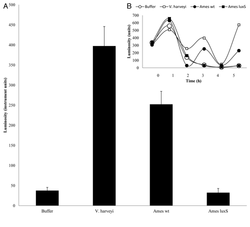
We created the B. anthracis Ames luxS mutant to eliminate endogenous production of the AI-2 reporter molecule and evaluate the effect of AI-2-deficient bacteria on mouse and guinea pig survival. Therefore, to show that we successfully eliminated AI-2 signal molecule production in the luxS mutant, we used filtered conditioned medium from wild-type and mutant B. anthracis cultures to induce bioluminescence in the V. harveyi BB170 reporter strain.Citation21 Filtered medium collected from stationary phase cultures of both the wild-type and luxS bacteria were added to the reporter strain and luminescence measured. The conditioned medium from the wild-type strain was able to induce bioluminescence, thus demonstrating the production of a functional AI-2 molecule in the Ames strain. However, when conditioned medium from the Ames luxS mutant was added to the reporter strain, the bioluminescence detected was no greater than what was observed with buffer alone (). These data confirm that the AI-2 signal molecule is not produced by the Ames luxS mutant.
Figure 1. Conditioned medium from the B. anthracis luxS mutant does not induce a V. harveyi QS response. AI-2 activity is shown as luminosity expressed from V. harveyi strain BB170 during growth in response to the addition of autoinducer buffer or filtered, conditioned medium from V. harveyi (BB170), the wild-type B. anthracis strain (Ames wt), or the B. anthracis luxS mutant (Ames luxS). (A) Luminosity values at 3 h of growth. The error bars represent the standard deviation from the readings of six replicates. The difference between the wild-type Ames and luxS mutant was statistically significant (p = 3.15 × 10⁻⁶). (B) Luminosity values at various growth times of the assay. These data are representative of the results of two independent experiments.

The luxS mutant is deficient in growth and delayed in sporulation
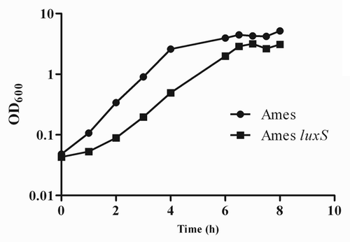
A previous report indicated that a luxS mutation in the attenuated Sterne strain of B. anthracis led to decreased growth in liquid culture.Citation13 Given this relationship between LuxS activity and bacterial growth, we compared the growth kinetics of the Ames wild-type and luxS mutant strains in brain heart infusion (BHI) broth. Consistent with the findings for B. anthracis Sterne, the Ames luxS mutant grew slower than wild-type Ames in liquid culture ().
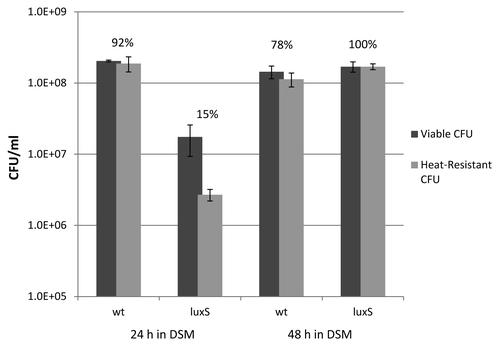
We also found that the luxS mutant was impaired for growth in Difco Sporulation Medium (DSM) when compared with the parental strain (data not shown) and measured about a log reduction in the total number of colony forming units (CFU)/ml at the 24 h time point (). While the drop in measured CFU/ml is likely due in part to slower growth, it may also be attributable to the more extensive aggregation of vegetative bacilli in luxS cultures than in wild-type cultures, which exhibited more free-floating spores under a light microscope (data not shown). Although luxS mutant was impaired for growth in DSM, the time at which the culture left exponential growth and began to sporulate was similar to that of the wild-type bacteria.
Figure 2. The B. anthracis luxS mutant exhibited impaired growth in BHI broth. Comparison is made of growth between the Ames wild-type (circles) and the luxS (squares) bacteria in BHI broth. These data are representative of the results of at least three independent experiments.

To confirm that the luxS mutant was producing fewer spores than wild-type bacteria, we analyzed sporulation by measuring the number of heat-resistant spores at 24 and 48 h after the onset of sporulation. Heat-resistance appears at an intermediate point in sporulation and is a reliable measure of progression through all but the final stages of spore formation.Citation22 At 24 h, only 15% of the viable luxS mutant cells were heat-resistant compared with 92% of wild-type bacteria. However, by 48 h, 100% of the luxS cells were resistant to high temperature (). These data suggest that while the luxS mutant is not significantly defective in spore production, it is defective in the progression through at least the early stages of the process. As a result, luxS cells progress through sporulation less efficiently or less rapidly than wild type.
Figure 3.LuxS-deficient bacteria show signs of delayed sporulation. Ames wild-type and luxS bacteria were inoculated at equal concentrations (by OD600) in DSM. CFU counts were obtained at 24 h and 48 h by plating culture samples before and after heating for 30 min at 65°C. The percentages of heat-resistant spores at each time point are indicated above the colony counts and are based on the fraction of heat-resistant cells in the sampled media. These data are representative of three independent experiments.

Finally, to assess the stability of luxS cointegrate mutants, 200 colonies from the sporulation assay were picked and patched onto Luria-Bertani (LB) agar plates with and without erythromycin. No erythromycin-sensitive colonies were detected, indicating that the cointegrate mutant was genetically stable during the in vitro culture conditions. We further validated mutant stability by PCR analysis of DNA derived from several of the antibiotic-resistant colonies. PCR analysis indicated that the luxS gene in each of the sampled colonies were disrupted by a pEO3 integrant (data not shown).
LuxS-deficient mutants remain as virulent as wild-type bacteria in mouse and guinea pig models
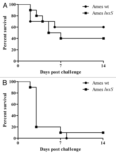
Because QS has been linked to virulence gene expression in many pathogens, including toxin gene expression in B. anthracis,Citation9,Citation13 we evaluated the Ames luxS mutant in multiple small animal models of anthrax infection. First, we compared survival of BALB/c mice challenged with either wild-type or luxS spores via intranasal (~2.65 × 10⁶ spores in 50 µl of water) or intraperitoneal (~2,700 spores in 100 µl of water) delivery. For these challenge models, previous studies calculated the Ames strain LD50 for mice to be 3.7 × 10⁴ and 500 spores, respectively.Citation23,Citation24 As shown in , there were no statistically significant differences in survival or time to death between mice challenged with wild-type or luxS mutant bacteria, regardless of challenge route.
Figure 4. Loss of LuxS AI-2 synthase activity does not affect B. anthracis virulence in mice. There was no statistical difference between the survival curves for BALB/c mice challenged with B. anthracis Ames luxS spores (squares; n = 10) or wild-type spores (circles; n = 10) regardless of whether the spores were administered intranasally (A) (~2.65 × 10⁶ spores; p = 0.72) or intraperitonally (B) (~2,700 spores; p = 0.49).

In addition to the mouse models of infection, guinea pigs (n = 10 for luxS and n = 5 for Ames wild-type) were challenged intramuscularly (~350 spores in 200 µl of water). A previous study calculated the Ames strain LD50 for a guinea pig to be ~100 spores.Citation25 Our study used a relatively low challenge dose so as to avoid overwhelming the animals and to ensure that a low level of attenuation in the virulence of spores could be observed. However, all animals similarly succumbed to infection with either strain by day 3, and no differences in survival or time to death were observed (data not shown).
Finally, to exclude the possibility that reversion of the luxS mutant to the wild-type form could be occurring during infection, guinea pig spleens were removed and bacteria recovered. A representative sample of bacteria (100 CFU from each mutant-challenged animal) was screened on LB agar plates with and without erythromycin. All plated CFU remained antibiotic resistant, indicating that the guinea pigs had succumbed to infection by luxS mutants and not wild-type revertants.
Discussion
Many bacterial species possess density- and growth phase-dependent genes, which respond to extracellular signaling molecules that accumulate in the environment. These QS signals regulate various bacterial functions and have been shown to affect the expression of numerous virulence factors.Citation8-Citation12 B. anthracis contains a functional AI-2 system that regulates the gene encoding the S-layer protein as well as other virulence genes, such as pagA, pagR, lef and cya.Citation8,Citation9,Citation13 These findings suggest that the AI-2 QS system might provide an effective therapeutic target against B. anthracis infection. To further evaluate this possibility, we constructed a LuxS-deficient mutant of the fully virulent B. anthracis Ames strain to determine if the LuxS AI-2 synthase is necessary for virulence in mouse and guinea pig models of infection.
The B. anthracis Ames luxS mutant exhibited an in vitro growth defect () similar to that previously observed in a luxS-deficient Sterne strain.Citation8 Additionally, a temporary delay was observed for completion of sporulation with the luxS mutant when compared with the parental strain (). To date, a role for the LuxS AI-2 synthase protein in B. anthracis sporulation has not been demonstrated. Furthermore, a recent microarray study, which compared the parental and luxS mutant Sterne strains,Citation13 did not reveal expression differences of known sporulation genes. However, a luxS mutant in the related Bacillus subtilis natto strain exhibited delayed formation of fruiting bodies during spore development.Citation26 B. subtilis sporulation is regulated by both the ComX peptide autoinducerCitation27 and small RNAs (sRNA).Citation28 Because AI-2 and sRNA are synergistic in regulating Vibrio choleraCitation29 and possibly B. anthracisCitation13 toxin expression, perhaps such combined regulation may explain the delayed sporulation results observed with the luxS mutant of B. anthracis.
The main goal of this study was to determine if the AI-2 QS pathway is significant for B. anthracis virulence. Therefore, we created and then evaluated virulence of an Ames luxS mutant in several small animal models for B. anthracis infection. BALB/c mice were tested by both intranasal and intraperitoneal routes of infection. In addition, guinea pigs were challenged intramuscularly. Despite previous studies linking AI-2 signal molecules to toxin and virulence factor regulation and the fact that our B. anthracis luxS mutant was deficient in growth, we saw no statistically significant differences in either the time to death or survival rates for animals challenged with mutant and wild-type isolates (). These results demonstrate that the intact AI-2 QS pathway does not play a significant role in B. anthracis infection in these animals and suggest that the AI-2 synthase enzyme would not provide an effective therapeutic target. Although LuxS activity is known to affect virulence in other bacterial species,Citation30-Citation32 our study using these rodent models of anthrax infection demonstrates that B. anthracis may be among those bacteria for which the AI-2 synthase does not play a significant role in infection.Citation33-Citation36
Methods
Bacterial growth and sporulation
Escherichia coli were cultured in LB medium supplemented with 50 μg/ml of ampicillin. The Ames strain of B. anthracisCitation37 was cultured in either LB medium or BHI broth. For construction and selection of the luxS mutant, medium was supplemented with 5 μg/ml of erythromycin, otherwise the mutant was grown under the same conditions as the wild-type strain. To induce sporulation, B. anthracis was grown in DSMCitation38 and spores were purified as previously described.Citation16 Because B. anthracis spores are resistant to heat, spore formation was measured by counting CFUs before and after incubating the bacterial cultures at 65°C for 30 min.
Mutant construction
The luxS mutant was constructed by PCR-amplifying an internal fragment of the B. anthracis Ames luxS gene (nucleotides 120–224, where ATG = 1–3) using two internal primers (5′-TTGCCAACCGAATAAAC-3′ and 5′-TCAAAATGTGGATAACGAT-3′) and cloning the PCR product into the pEO3 plasmid.Citation19 The resulting construct was integrated into the B. anthracis Ames chromosome to form a merodiploid mutant as previously described.Citation16 PCR analysis was used to confirm stabile disruption of the luxS gene in both culture- and animal-passaged bacteria.
AI-2 production
A previously published bioluminescence assay was used, with minor modifications, to detect endogenously produced AI-2 signal molecules in wild-type and luxS B. anthracis cultures.Citation21 Briefly, an overnight culture of V. harveyi strain BB170 (ATCC) was grown in LB at 30°C, diluted 1:5,000 into autoinducer buffer, and 990 µl aliquots were distributed into optical-grade micro-titer plates preloaded with 10 µl of conditioned medium. The filtered, conditioned medium was prepared from liquid bacterial cultures of B. anthracis Ames wild-type or luxS strains grown to stationary phase in BHI at 37°C. Similarly prepared filtered medium from V. harveyi was used as a control. The assay plate was incubated at 30°C with orbital shaking, and luminescence was measured at 490 nm every hour using a Victor2 Multilabel Counter (Perkin-Elmer). Each test subject was averaged from six replicates across a single microtiter plate.
Animal challenges
To assess potential changes in virulence associated with disrupting the B. anthracis Ames AI-2 QS pathway, spores from both wild-type and mutant strains were used in mouse intraperitoneal and intranasal models,Citation39 as well as the guinea pig intramuscular model.Citation25 Research was conducted under an IACUC-approved protocol in compliance with the Animal Welfare Act, PHS Policy and other federal statutes and regulations relating to animals and experiments involving animals. The facility where this research was conducted is accredited by the Association for Assessment and Accreditation of Laboratory Animal Care, International and adheres to principles stated in the Guide for the Care and Use of Laboratory Animals, National Research Council, 2011.
Statistics
Survival rates were compared between groups by Fisher exact tests with permutation adjustment for multiple comparisons using SAS Version 8.2 (SAS Institute Inc., SAS OnlineDoc, Version 8). For comparing data from bioluminescence experiments and time to death studies of mouse challenges, statistical significance (p < 0.05) was determined by the two-tailed Student’s t-test.
Acknowledgments
We thank Gabriel Rother for his invaluable technical assistance, Diane Fisher for completing the statistical analysis and Adam Driks and Brad Stiles for their helpful comments and review of this manuscript. The research described herein was sponsored by the Defense Threat Reduction Agency JSTO-CBD project 1.1A0021_07_RD_B. Opinions, interpretations, conclusions and recommendations are those of the authors and are not necessarily endorsed by the United States Army.
Disclosure of Potential Conflicts of Interest
No potential conflicts of interest were disclosed.
References
- Friedlander AM. Anthrax: clinical features, pathogenesis, and potential biological warfare threat. Curr Clin Top Infect Dis 2000; 20:335 - 49; PMID: 10943532
- Driks A. The dynamic spore. Proc Natl Acad Sci U S A 2003; 100:3007 - 9; http://dx.doi.org/10.1073/pnas.0730807100; PMID: 12631692
- Moir A, Corfe BM, Behravan J. Spore germination. Cell Mol Life Sci 2002; 59:403 - 9; http://dx.doi.org/10.1007/s00018-002-8432-8; PMID: 11964118
- Brossier F, Mock M. Toxins of Bacillus anthracis. Toxicon 2001; 39:1747 - 55; http://dx.doi.org/10.1016/S0041-0101(01)00161-1; PMID: 11595637
- Inglesby TV, Henderson DA, Bartlett JG, Ascher MS, Eitzen E, Friedlander AM, et al, Working Group on Civilian Biodefense. Anthrax as a biological weapon: medical and public health management. [see comments] [published erratum appears in JAMA 2000 Apr 19;283(15):1963] JAMA 1999; 281:1735 - 45; http://dx.doi.org/10.1001/jama.281.18.1735; PMID: 10328075
- Winzer K, Hardie KR, Burgess N, Doherty N, Kirke D, Holden MT, et al. LuxS: its role in central metabolism and the in vitro synthesis of 4-hydroxy-5-methyl-3(2H)-furanone. Microbiology 2002; 148:909 - 22; PMID: 11932438
- Tu KC, Waters CM, Svenningsen SL, Bassler BL. A small-RNA-mediated negative feedback loop controls quorum-sensing dynamics in Vibrio harveyi. Mol Microbiol 2008; 70:896 - 907; PMID: 18808382
- Jones MB, Blaser MJ. Detection of a luxS-signaling molecule in Bacillus anthracis. Infect Immun 2003; 71:3914 - 9; http://dx.doi.org/10.1128/IAI.71.7.3914-3919.2003; PMID: 12819077
- Jones MB, Jani R, Ren D, Wood TK, Blaser MJ. Inhibition of Bacillus anthracis growth and virulence-gene expression by inhibitors of quorum-sensing. J Infect Dis 2005; 191:1881 - 8; http://dx.doi.org/10.1086/429696; PMID: 15871122
- Siller M, Janapatla RP, Pirzada ZA, Hassler C, Zinkl D, Charpentier E. Functional analysis of the group A streptococcal luxS/AI-2 system in metabolism, adaptation to stress and interaction with host cells. BMC Microbiol 2008; 8:188; http://dx.doi.org/10.1186/1471-2180-8-188; PMID: 18973658
- Burgess NA, Kirke DF, Williams P, Winzer K, Hardie KR, Meyers NL, et al. LuxS-dependent quorum sensing in Porphyromonas gingivalis modulates protease and haemagglutinin activities but is not essential for virulence. Microbiology 2002; 148:763 - 72; PMID: 11882711
- Marouni MJ, Sela S. The luxS gene of Streptococcus pyogenes regulates expression of genes that affect internalization by epithelial cells. Infect Immun 2003; 71:5633 - 9; http://dx.doi.org/10.1128/IAI.71.10.5633-5639.2003; PMID: 14500483
- Jones MB, Peterson SN, Benn R, Braisted JC, Jarrahi B, Shatzkes K, et al. Role of luxS in Bacillus anthracis growth and virulence factor expression. Virulence 2010; 1:72 - 83; http://dx.doi.org/10.4161/viru.1.2.10752; PMID: 21178420
- Jones MB, Blaser MJ. Detection of a luxS-signaling molecule in Bacillus anthracis. Infect Immun 2003; 71:3914 - 9; http://dx.doi.org/10.1128/IAI.71.7.3914-3919.2003; PMID: 12819077
- Read TD, Peterson SN, Tourasse N, Baillie LW, Paulsen IT, Nelson KE, et al. The genome sequence of Bacillus anthracis Ames and comparison to closely related bacteria. Nature 2003; 423:81 - 6; http://dx.doi.org/10.1038/nature01586; PMID: 12721629
- Bozue JA, Parthasarathy N, Phillips LR, Cote CK, Fellows PF, Mendelson I, et al. Construction of a rhamnose mutation in Bacillus anthracis affects adherence to macrophages but not virulence in guinea pigs. Microb Pathog 2005; 38:1 - 12; http://dx.doi.org/10.1016/j.micpath.2004.10.001; PMID: 15652290
- Giorno R, Bozue J, Cote C, Wenzel T, Moody KS, Mallozzi M, et al. Morphogenesis of the Bacillus anthracis spore. J Bacteriol 2007; 189:691 - 705; http://dx.doi.org/10.1128/JB.00921-06; PMID: 17114257
- Giorno R, Mallozzi M, Bozue J, Moody KS, Slack A, Qiu D, et al. Localization and assembly of proteins comprising the outer structures of the Bacillus anthracis spore. Microbiology 2009; 155:1133 - 45; http://dx.doi.org/10.1099/mic.0.023333-0; PMID: 19332815
- Mendelson I, Tobery S, Scorpio A, Bozue J, Shafferman A, Friedlander AM. The NheA component of the non-hemolytic enterotoxin of Bacillus cereus is produced by Bacillus anthracis but is not required for virulence. Microb Pathog 2004; 37:149 - 54; http://dx.doi.org/10.1016/j.micpath.2004.06.008; PMID: 15351038
- Welkos S, Friedlander A, Weeks S, Little S, Mendelson I. In-vitro characterisation of the phagocytosis and fate of anthrax spores in macrophages and the effects of anti-PA antibody. J Med Microbiol 2002; 51:821 - 31; PMID: 12435060
- Taga ME. Methods for analysis of bacterial autoinducer-2 production. Curr Protoc Microbiol 2005; Chapter 1:Unit 1C -8.
- Cutting SM, Vander Horn PB. Molecular biological methods for Bacillus. Chichester, United Kingdom: John Wiley & Sons Ltd., 1990.
- Lyons CR, Lovchik J, Hutt J, Lipscomb MF, Wang E, Heninger S, et al. Murine model of pulmonary anthrax: kinetics of dissemination, histopathology, and mouse strain susceptibility. Infect Immun 2004; 72:4801 - 9; http://dx.doi.org/10.1128/IAI.72.8.4801-4809.2004; PMID: 15271942
- Popov SG, Popova TG, Grene E, Klotz F, Cardwell J, Bradburne C, et al. Systemic cytokine response in murine anthrax. Cell Microbiol 2004; 6:225 - 33; http://dx.doi.org/10.1046/j.1462-5822.2003.00358.x; PMID: 14764106
- Fellows PF, Linscott MK, Ivins BE, Pitt ML, Rossi CA, Gibbs PH, et al. Efficacy of a human anthrax vaccine in guinea pigs, rabbits, and rhesus macaques against challenge by Bacillus anthracis isolates of diverse geographical origin. Vaccine 2001; 19:3241 - 7; http://dx.doi.org/10.1016/S0264-410X(01)00021-4; PMID: 11312020
- Lombardía E, Rovetto AJ, Arabolaza AL, Grau RR. A LuxS-dependent cell-to-cell language regulates social behavior and development in Bacillus subtilis. J Bacteriol 2006; 188:4442 - 52; http://dx.doi.org/10.1128/JB.00165-06; PMID: 16740951
- Waters CM, Bassler BL. Quorum sensing: cell-to-cell communication in bacteria. Annu Rev Cell Dev Biol 2005; 21:319 - 46; http://dx.doi.org/10.1146/annurev.cellbio.21.012704.131001; PMID: 16212498
- Silvaggi JM, Perkins JB, Losick R. Genes for small, noncoding RNAs under sporulation control in Bacillus subtilis. J Bacteriol 2006; 188:532 - 41; http://dx.doi.org/10.1128/JB.188.2.532-541.2006; PMID: 16385044
- Miller MB, Skorupski K, Lenz DH, Taylor RK, Bassler BL. Parallel quorum sensing systems converge to regulate virulence in Vibrio cholerae. Cell 2002; 110:303 - 14; http://dx.doi.org/10.1016/S0092-8674(02)00829-2; PMID: 12176318
- Labandeira-Rey M, Janowicz DM, Blick RJ, Fortney KR, Zwickl B, Katz BP, et al. Inactivation of the Haemophilus ducreyi luxS gene affects the virulence of this pathogen in human subjects. J Infect Dis 2009; 200:409 - 16; http://dx.doi.org/10.1086/600142; PMID: 19552526
- Stroeher UH, Paton AW, Ogunniyi AD, Paton JC. Mutation of luxS of Streptococcus pneumoniae affects virulence in a mouse model. Infect Immun 2003; 71:3206 - 12; http://dx.doi.org/10.1128/IAI.71.6.3206-3212.2003; PMID: 12761100
- Winzer K, Sun YH, Green A, Delory M, Blackley D, Hardie KR, et al. Role of Neisseria meningitidis luxS in cell-to-cell signaling and bacteremic infection. Infect Immun 2002; 70:2245 - 8; http://dx.doi.org/10.1128/IAI.70.4.2245-2248.2002; PMID: 11895997
- Perrett CA, Karavolos MH, Humphrey S, Mastroeni P, Martinez-Argudo I, Spencer H, et al. LuxS-based quorum sensing does not affect the ability of Salmonella enterica serovar Typhimurium to express the SPI-1 type 3 secretion system, induce membrane ruffles, or invade epithelial cells. J Bacteriol 2009; 191:7253 - 9; http://dx.doi.org/10.1128/JB.00727-09; PMID: 19783624
- Zhu C, Feng S, Sperandio V, Yang Z, Thate TE, Kaper JB, et al. The possible influence of LuxS in the in vivo virulence of rabbit enteropathogenic Escherichia coli. Vet Microbiol 2007; 125:313 - 22; http://dx.doi.org/10.1016/j.vetmic.2007.05.030; PMID: 17643872
- Blevins JS, Revel AT, Caimano MJ, Yang XF, Richardson JA, Hagman KE, et al. The luxS gene is not required for Borrelia burgdorferi tick colonization, transmission to a mammalian host, or induction of disease. Infect Immun 2004; 72:4864 - 7; http://dx.doi.org/10.1128/IAI.72.8.4864-4867.2004; PMID: 15271949
- Jordan DM, Sperandio V, Kaper JB, Dean-Nystrom EA, Moon HW. Colonization of gnotobiotic piglets by a luxS mutant strain of Escherichia coli O157:H7. Infect Immun 2005; 73:1214 - 6; http://dx.doi.org/10.1128/IAI.73.2.1214-1216.2005; PMID: 15664967
- Little SF, Knudson GB. Comparative efficacy of Bacillus anthracis live spore vaccine and protective antigen vaccine against anthrax in the guinea pig. Infect Immun 1986; 52:509 - 12; PMID: 3084385
- Schaeffer P, Millet J, Aubert JP. Catabolic repression of bacterial sporulation. Proc Natl Acad Sci U S A 1965; 54:704 - 11; http://dx.doi.org/10.1073/pnas.54.3.704; PMID: 4956288
- Cote CK, Rea KM, Norris SL, van Rooijen N, Welkos SL. The use of a model of in vivo macrophage depletion to study the role of macrophages during infection with Bacillus anthracis spores. Microb Pathog 2004; 37:169 - 75; http://dx.doi.org/10.1016/j.micpath.2004.06.013; PMID: 15458777