
Figures & data
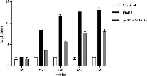
New Zealand rabbits were immunized with 250 μg of the pcDNA3/FlaB3 plasmid, 150 μg of pcDNA3 as a negative control or 150 μg of recombinant flagellin as a positive control. The serum IgG levels were analyzed by ELISA. The means and standard derivations for the antibody levels were calculated from three rabbits
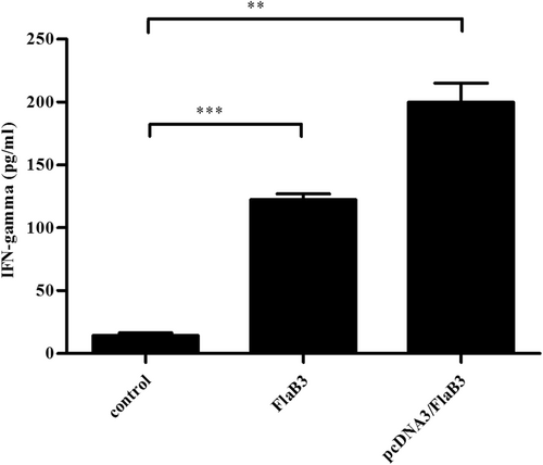
Sera collected from immunized and nonimmunized rabbits were evaluated with an IFN-γ ELISA kit according to the manufacturer’s protocols. The means ± SDs are the results from three individual rabbit sera in three independent experiments (**P < 0.01, ***P < 0.001)
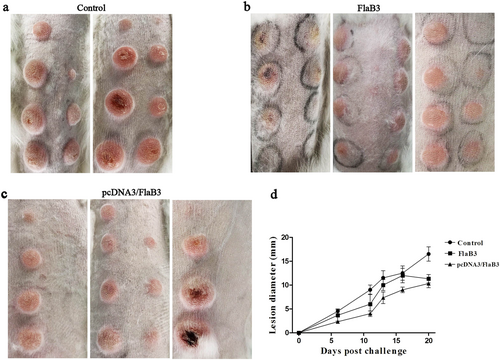
The pcDNA3/FlaB3-immunized rabbits had fewer ulcerative lesions than the unimmunized controls and the lesion diameter measured in the control animals was larger than that in immunized animals within 21 days postinfection (a, b, c). Eight rabbits were collected on day 21 post-intradermal challenge with the T. pallidum Nichols strain at eight locations on their backs. Skin lesions were observed and measured at the challenged sites every 3 days (d). The results are presented as the median ± 95% confidence interval between individual immunized or control animals. Significance was assessed using nonlinear regression and the extra sum of squares F-test (P < 0.05 (d))
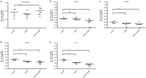
The numbers of T. pallidum in the lesions
The T. pallidum burden was evaluated in control animals (N = 2) and animals immunized with pcDNA3/FlaB3 (N = 3) or recombinant flagellin (N = 3) using quantitative real-time PCR to measure the flaA DNA concentrations in the lesion biopsies ((a); lesion biopsy) and disseminated infection sites ((b) blood, (c) testicles, (d) spleen, and (e) liver). The results were normalized to the rabbit gDNA concentrations using the Mann–Whitney test. The points correspond to three samples that were separately extracted from each animal. Horizontal lines represent mean values (*P < 0.05, **P < 0.01, ***P < 0.001, NS nonsignificant)
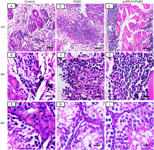
Inflammatory infiltrates in pcDNA3/FlaB3- and FlaB3-immunized rabbits display a dense diffuse pattern (b, c) consisting predominantly of lymphocytes, macrophages, and plasma cells (solid arrow) (e, f). Interstitial inflammation of the testicular tissues is localized and predominantly lymphohistiocytic (solid arrow = lymphocyte; red arrow = macrophage; dashed arrow = plasma cell) in the control group (g) but not in the immunized rabbits (h, i). Skin and testicles of pcDNA3/FlaB3-, FlaB3-, and pcDNA3-immunized rabbits were sectioned and stained with H&E on day 21 after T. pallidum infection. Rabbits immunized with pcDNA3 were used as the controls (a)