Figures & data
Figure 1. Stratigraphy of cetacean-bearing units relevant to fossil dolphin ZMT 73. For international occurrences, only the key Early Miocene formations are indicated; see text for details. For New Zealand, the key zonal species of foraminifera for Oligocene to Early Miocene stages are based on Hornibrook et al. (Citation1989) and references discussed in the text. Schematic lithostratigraphy for the southern Canterbury Basin is based on Hornibrook et al. (Citation1989), modified by Fordyce’s field observations of vertebrate-bearing strata. The selected species of Odontoceti are shown in approximate age for the Canterbury Basin formations, although ZMT 73 and Papahu taitapu are from other sequences, as discussed in the text.
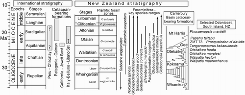
Figure 3. Skull materials of ZMT 73. A, Dorsal view. B, Ventral view (see for detailed basicranium). C, Left lateral view. D, Posterior view.
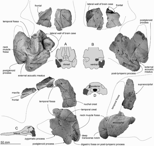
Figure 4. Details of partial left basicranium of ZMT 73. Ventral view.
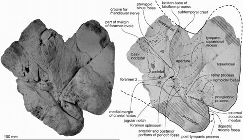
Figure 5. Teeth of ZMT 73. A–D, One complete tooth. E–G, Three crowns lacking roots. H–J, Crown with a denticle. A, Anterior view. B, Posterior view. C, Buccal view. D, Lingual view. E–I, Buccal or lingual views. J, Distal (apical) view.
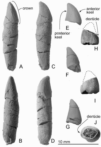
Figure 6. Right periotic of ZMT 73. A, Ventral view. B, Dorsomedial view showing foramina in the internal acoustic meatus. C, Lateral view. D, Medial view. E, Posterior view. F, Dorsal view. G, Anterior view.
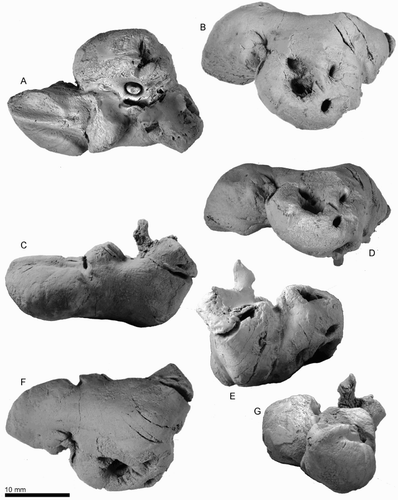
Figure 7. Key features of the right periotic of ZMT 73. A, Ventral view. B, Dorsomedial view showing foramina in the internal acoustic meatus. C, Lateral view. D, Medial view. E, Posterior view. F, Dorsal view. G, Anterior view.
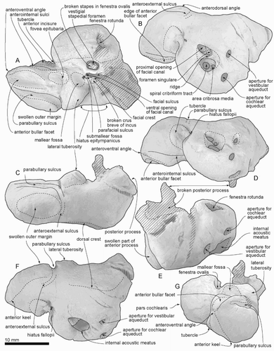
Figure 8. Right bulla of ZMT 73. A, Dorsal view. B, Lateral view. C, Posterior view. D, Medial view. E, Ventral view. F, Anterior view.
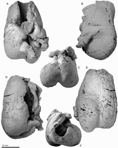
Figure 9. Key features of the right bulla of ZMT 73. A, Dorsal view. B, Lateral view. C, Posterior view. D, Medial view. E, Ventral view. F, Anterior view.
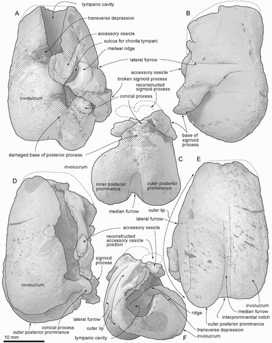
Figure 10. Incus and malleus of ZMT 73. A, Lateral view. B, Dorsal view. C, Anterior view. D, Posterior view. E, Medial view. F, Ventral view.
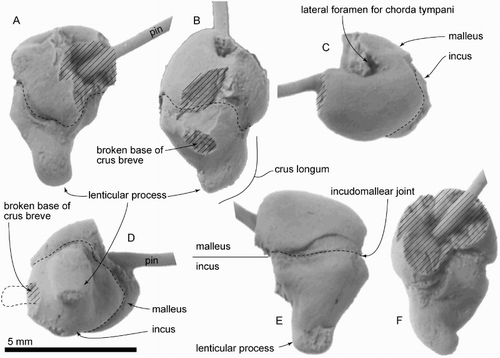
Figure 11. Provisionally identified supraoccipital and rostrum of ZMT 73. A, B, Part of the putative supraoccipital. C–E, Part of the putative rostrum. C, Putative rostral elements in original preserved contact. D, E, Putative rostral elements separated, showing both sides.
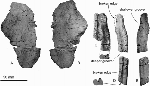
Figure 2. Locality map and stratigraphy, based on Rattenbury et al. (Citation2006).
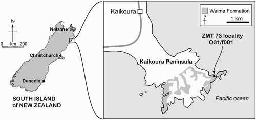
Table 1. Measurements in mm of ZMT 73: periotic and tympanic bulla. Dimensions follow Tanaka & Fordyce (2015a).
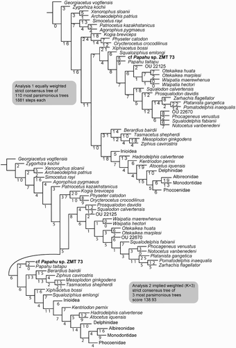
Figure 12. Phylogenetic analysis of ZMT 73 and Papahu taitapu. The clades Inioidea, Phocoenidae and Delphinidae are collapsed. (Cladograms with all clades shown are in Figures S1 and S2.) Top, Strict consensus tree of equally weighted Analysis 1 with branch length labelled. Bottom, Strict consensus tree of implied weighting Analysis 2 with branch length labelled.