730
Views
0
CrossRef citations to date
0
Altmetric
Research articles

Early Miocene brachiopods of the Waitemata and Waitakere groups, Auckland and Northland, New Zealand

Pages 2-22 | Received 06 Oct 2015, Accepted 19 Sep 2016, Published online: 13 Oct 2016

Figures & data

Table 1. Fossil brachiopod specimens from the Waitemata Group strata collected as part of the Novara Expedition (Flügel 1959). These are held in the Natural History Museum, Vienna, Austria.

Table 2. The groups, subgroups and formations mentioned in this study.

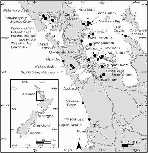
Figure 1. Localities mentioned in this study.

Figure 1. Localities mentioned in this study.

Table 3. Latitude and longitude for localities in Figure 1.

Table 4. Material examined in this study.

Table 5. Material examined in this study.

Figure 2. A–D, Notosaria antipoda. A, MA34672a. Dorsal exterior. B, B629. Dorsal exterior. C–D, MA34672b. C, Dorsal interior. D, Close up of sockets, socket ridges and septum. E, F, Thecidellina cf. maxilla CM 2006.64.92. E, Dorsal interior. F, Dorsal interior, posterior tilted forward. GI, Liothyrella gravida. Type specimens. G, H, NHMW‐1959-0335-0012a. G, Ventral exterior. H, Lateral exterior (ventral valve only). I, NHMW‐1865-0037-0105a. Dorsal exterior of partial specimen. JN, Terebratulina suessi. J, OU45764. Dorsal exterior of juvenile. K–N, MA77312. K, Dorsal exterior. L, Ventral exterior. M, Lateral exterior. N, Anterior commissure. E–G photographed by Alice Schumacher of the NHMW. All scale bars 5 mm unless indicated otherwise. se, septum; sp, broken spine base. B = University of Auckland School of Environment, CM = Canterbury Museum, Christchurch, MA = Auckland Museum, NHMW = Naturhistorisches Museum Vienna, OU = University of Otago Geology Museum, Dunedin.
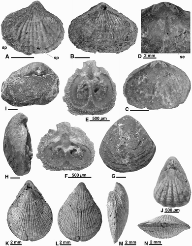
Figure 2. A–D, Notosaria antipoda. A, MA34672a. Dorsal exterior. B, B629. Dorsal exterior. C–D, MA34672b. C, Dorsal interior. D, Close up of sockets, socket ridges and septum. E, F, Thecidellina cf. maxilla CM 2006.64.92. E, Dorsal interior. F, Dorsal interior, posterior tilted forward. G–I, Liothyrella gravida. Type specimens. G, H, NHMW‐1959-0335-0012a. G, Ventral exterior. H, Lateral exterior (ventral valve only). I, NHMW‐1865-0037-0105a. Dorsal exterior of partial specimen. J–N, Terebratulina suessi. J, OU45764. Dorsal exterior of juvenile. K–N, MA77312. K, Dorsal exterior. L, Ventral exterior. M, Lateral exterior. N, Anterior commissure. E–G photographed by Alice Schumacher of the NHMW. All scale bars 5 mm unless indicated otherwise. se, septum; sp, broken spine base. B = University of Auckland School of Environment, CM = Canterbury Museum, Christchurch, MA = Auckland Museum, NHMW = Naturhistorisches Museum Vienna, OU = University of Otago Geology Museum, Dunedin.

Figure 3. AK, Magasella neozelandica. A, NHMW‐1865-0037-0150i. Dorsal exterior. B, MA91104a. Dorsal exterior. C, D, B630. C, Dorsal exterior. D, Lateral exterior. E, NHMW‐1865-0037-0150b. Anterior commissure. F, MA91104b. Anterior commissure. G, B631. Ventral exterior. H, I, B632. H, Strong beak ridges, disjunct deltidial plates. I, Erect beak. J, B633. Cardinalia. K, B634. Cardinalia and septum. LO, Magasella sanguinea. L–N, OU44534a. L, Dorsal exterior. M, Anterior commissure. N, Less robust beak ridges, conjunct deltidial plates. O, OU44534b. Cardinalia and septum. A and E photographed by Alice Schumacher of the NHMW. All scale bars 5 mm unless indicated otherwise. B = University of Auckland School of Environment, MA = Auckland Museum, NHMW = Naturhistorisches Museum Vienna.
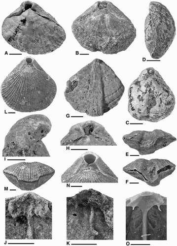
Figure 3. A–K, Magasella neozelandica. A, NHMW‐1865-0037-0150i. Dorsal exterior. B, MA91104a. Dorsal exterior. C, D, B630. C, Dorsal exterior. D, Lateral exterior. E, NHMW‐1865-0037-0150b. Anterior commissure. F, MA91104b. Anterior commissure. G, B631. Ventral exterior. H, I, B632. H, Strong beak ridges, disjunct deltidial plates. I, Erect beak. J, B633. Cardinalia. K, B634. Cardinalia and septum. L–O, Magasella sanguinea. L–N, OU44534a. L, Dorsal exterior. M, Anterior commissure. N, Less robust beak ridges, conjunct deltidial plates. O, OU44534b. Cardinalia and septum. A and E photographed by Alice Schumacher of the NHMW. All scale bars 5 mm unless indicated otherwise. B = University of Auckland School of Environment, MA = Auckland Museum, NHMW = Naturhistorisches Museum Vienna.

Figure 4. AG. Magadina browni. A, B635. Juvenile dorsal exterior. B, B636. Adult dorsal exterior. C, B637. Lateral exterior. D, B638. Ventral interior. E, B639. Juvenile dorsal interior. F, G, B640. F. Adult dorsal interior. G, Oblique view. All scale bars 2 mm. cp, cardinal process; gr, groove; sr, socket ridges; vs, ventral septum. B = University of Auckland School of Environment.
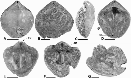
Figure 4. A–G. Magadina browni. A, B635. Juvenile dorsal exterior. B, B636. Adult dorsal exterior. C, B637. Lateral exterior. D, B638. Ventral interior. E, B639. Juvenile dorsal interior. F, G, B640. F. Adult dorsal interior. G, Oblique view. All scale bars 2 mm. cp, cardinal process; gr, groove; sr, socket ridges; vs, ventral septum. B = University of Auckland School of Environment.

Figure 5. AN, ‘Pachymagas’. A–D, MA91060a. Juvenile specimen. A, Dorsal exterior. B, Ventral exterior. C, Lateral exterior. D, Anterior commissure. E, MA91060b. Mid-sized specimen. Dorsal exterior. F–I, B641. Adult specimen. F, Dorsal exterior. G, Ventral exterior. H, Lateral exterior. I, Sulcate anterior commissure. J, B642. Cardinalia of partial dorsal valve of mid-sized specimen. K–N, Images from CT scan of MA91060b. Figure K is the most dorsal, L–N are progressively more ventral. O, B643. Swollen cardinalia of adult specimen. PR, OU54765a‐c. Terebratellidae indet. P, Whole juvenile specimen. Q, Partial ventral valve. R, Partial dorsal valve. All scale bars 5 mm unless indicated otherwise. cp, cardinal process; se, septum; slm, septalium; sr, socket ridge; thsr, thickened socket ridge. B = University of Auckland School of Environment, MA = Auckland Museum, OU = University of Otago Geology Museum, Dunedin.
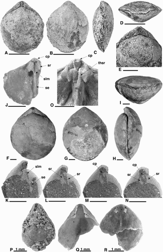
Figure 5. A–N, ‘Pachymagas’. A–D, MA91060a. Juvenile specimen. A, Dorsal exterior. B, Ventral exterior. C, Lateral exterior. D, Anterior commissure. E, MA91060b. Mid-sized specimen. Dorsal exterior. F–I, B641. Adult specimen. F, Dorsal exterior. G, Ventral exterior. H, Lateral exterior. I, Sulcate anterior commissure. J, B642. Cardinalia of partial dorsal valve of mid-sized specimen. K–N, Images from CT scan of MA91060b. Figure K is the most dorsal, L–N are progressively more ventral. O, B643. Swollen cardinalia of adult specimen. P–R, OU54765a‐c. Terebratellidae indet. P, Whole juvenile specimen. Q, Partial ventral valve. R, Partial dorsal valve. All scale bars 5 mm unless indicated otherwise. cp, cardinal process; se, septum; slm, septalium; sr, socket ridge; thsr, thickened socket ridge. B = University of Auckland School of Environment, MA = Auckland Museum, OU = University of Otago Geology Museum, Dunedin.

Reprints and Corporate Permissions

Please note: Selecting permissions does not provide access to the full text of the article, please see our help page How do I view content?

To request a reprint or corporate permissions for this article, please click on the relevant link below:

Academic Permissions

Please note: Selecting permissions does not provide access to the full text of the article, please see our help page How do I view content?

Obtain permissions instantly via Rightslink by clicking on the button below:

If you are unable to obtain permissions via Rightslink, please complete and submit this Permissions form. For more information, please visit our Permissions help page.