Figures & data
Table 1 Strains used in the present study and their probe reactivities for the HSIG 1451 probe.
Figure 1 Light and fluorescence micrographs of Heterosigma akashiwo strain HYM06HA. A, Live cell. B, Chlorophyll in the live cell. C, Cell fixed with paraformaldehyde (PFA). D, Chlorophyll in the fixed cell. E, Cell following chlorophyll extraction with methanol after PFA fixation. F, Nucleus stained with propidium iodide (PI) after methanol treatment. G, Cell following chlorophyll extraction with ethanol after PFA fixation. H, Nucleus stained with PI after ethanol treatment.
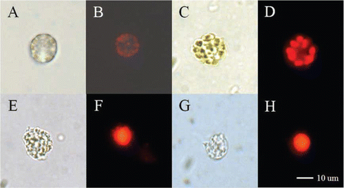
Table 2 Oligonucleotide probes used in the present study.
Table 3 Specificities of the HSIG1451 probe.
Figure 2 Effect of probe concentration on fluorescent intensity (arbitrary units, AU) in Heterosigma akashiwo cells.
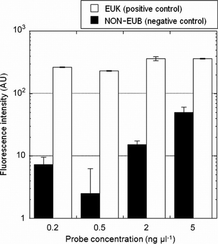
Figure 3 Effect of formamide concentration on fluorescent intensity (arbitrary units, AU) in Heterosigma akashiwo cells.
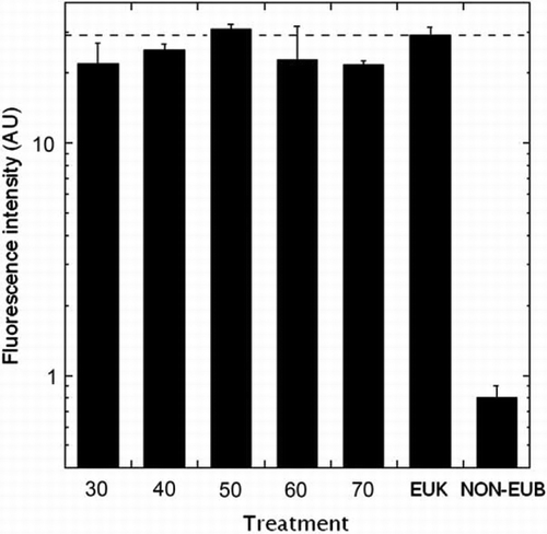
Figure 4 Epifluorescence micrographs. A, Algal cells after fluorescence in situ hybridisation using various probes. Panels a, f, and k: Heterosigma akashiwo, panels b, g and l: Chattonella sp., panels c and h: Gymnodinium impudicum, panels d and i: Akashiwo sanguine, panels e and j: Prorocentrum micans; upper panels (a, b, c, d, and e): cells hybridised with the EUK1209 probe, middle panels (f, g, h, I, and j): cells hybridised with the HSIG1451 probe, lower panels (k and l): cells hybridised with the NON338 probe. B, Natural seawater sample collected from Geoje Island. Panel a: cells hybridised with HSIG1451 probe, panel b: cells hybridised with EUK1209 probe.
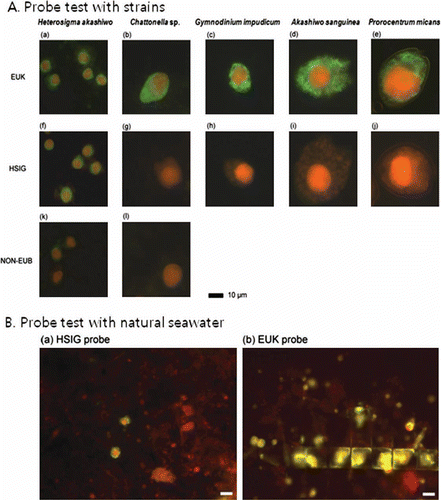
Figure 5 Flow cytometric signatures of Heterosigma akashiwo. A–C, Signatures of H. akashiwo strain after fluorescence in situ hybridization (FISH). D–I, Signatures of cells collected from Korean coastal waters after FISH. A, Detection of H. akashiwo cells after FISH; red dots indicate 6 m beads; green dots indicate H. akashiwo cells detected with green fluorescence after FISH. B, Discrimination of doublet and singlet cells of H. akashiwo; pink dots indicate singlet cells discriminated by peak area and peak width (length) of red fluorescence of detected cells (green dots) in panel A. C, DNA histograms of H. akashiwo gated in panel B (composed of singlet cells discriminated in panel B). D–F, Samples hybridised with HSIG1284 probe. G–I, Samples hybridised with NON338 probe.
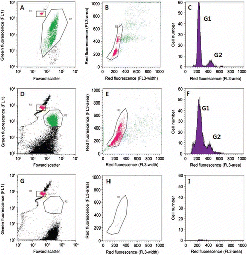
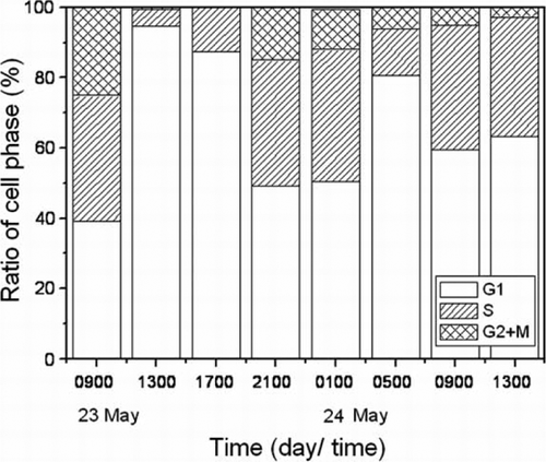
Figure 6 Diel cell cycle analysis of Heterosigma akashiwo from Shiwha Lake after fluorescence in situ hybridisation.