Figures & data
Table 1. Description and diagnostic morphologies of all currently known extant Palusphaera species vs other spine-bearing taxa of the family Rhabdosphaeraceae. SEM images are not to the same scale
Figs 1–4. Ultrastructure of Rhabdosphaeraceae
Table 2. List of all sampling localities, including physicochemical data, with Palusphaera specimens used in the present study
Fig. 5. Map showing the locations from which Palusphaera specimens were obtained
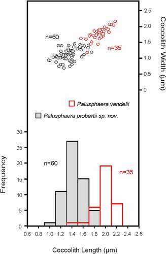
Fig. 6. Frequency and bivariate plots of the main morphological parameters (coccolith length and width) measured in specimens of Palusphaera vandelii (image codes 222-04, 289-62, 246-06a 262-20, 247-17 276-30, 280-14) and Palusphaera probertii sp. nov. (image codes 118-55, 188-03, 288-06, 297-77)
Figs 7–11. SEM micrographs of Palusphaera vandelii. Scale bar = 5 μm for Figs 7, 8, 9, 11
Figs 12–16. SEM micrographs of Palusphaera probertii sp. nov
Figs 17–21. SEM micrographs of Palusphaera crosiae sp. nov. Scale bar = 5 μm for Figs 17, 18, 19
Figs 22–24. SEM micrographs of Palusphaera bownii sp. nov. Scale bar = 2 μm
Fig. 25. Schematic representation of the coccolith morphology and ultrastructure of all currently known morphospecies in the genus Palusphaera