
Figures & data
Table 1. CO1 sequences used in this study.
Figure 1. Holotype of Siphonethus enotatus Chamberlin, Citation1920 (MCZ4885), photographs by Laura Leibensperger (MCZ). (a) Habitus, lateral view. (b) Head and anterior body-rings, ventral view. (c) Preanal ring and posterior body-rings, lateral view.
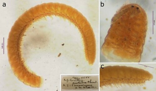
Figure 2. Holotype of Siphonethus bellus Chamberlin, Citation1920 (MCZ4878), photographs by Laura Leibensperger (MCZ). (a) Habitus, lateral view. (b) Head and anterior body-rings, lateral view. (c) Preanal ring and posterior body-rings, lateral view.
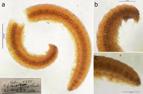
Table 2. Comparison of Siphonethus species. Information for S. enotatus and S. bellus taken from the original description (Chamberlin Citation1920) and inferred from photographs of types. *might be an artefacts due to preservation. Inverted commas (‘ … ’) indicate text directly quoted from Chamberlin (Citation1920: 99). Question marks (?) indicate unknown character states.
Figure 3. Habitat of Siphonethus dudleycookeorum sp. nov. in Lamorran HouseGardens (Great Britain, Cornwall), photographs by S. J. Gregory. (a) Path lined by planted tree ferns from New Zealand. (b) Siphonethus dudleycookeorum sp. nov.
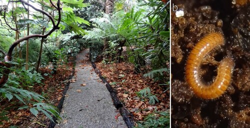
Figure 4. Siphonethus dudleycookeorum sp. nov. from Lamorran House Gardens (Great Britain, Cornwall), female. (a) Habitus, lateral view. (b) Anterior body rings, dorsal view. (c) Midbody-rings dorsal view.
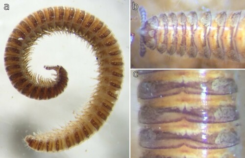
Figure 5. Siphonethus dudleycookeorum sp. nov., male holotype (ZFMK-MYR11376) from Great Britain, head, SEM images. (a) Head, frontal view. (b) Head, lateral view. (c) Detail of labrum. (d) Gnathochilarium. (e) Antennae, overview. (f) Antennae apical antennomeres, apical view. (g) Antennae apical antennomeres, lateral view. Scale: a, b, d, e = 100 µm, c, f = 10 µm, g = 20 µm. Abbreviations: I-VII = antennomere 1-7, ac = apical cones, at = antennae; co = collum, gn = gnathochilarium, in = incision of labrum, la = labrum, ms = macrosetae, om = ommatidia, sb = sensilla basiconica.
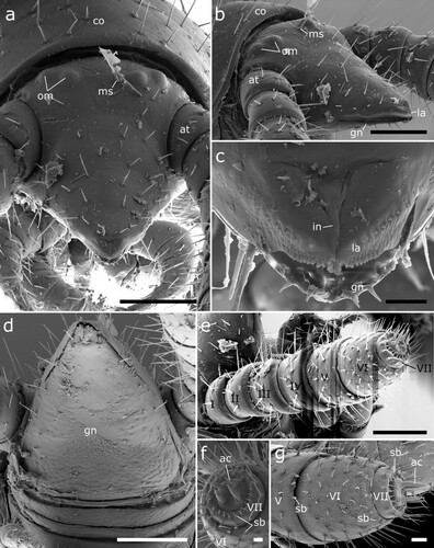
Figure 6. Siphonethus dudleycookeorum sp. nov., female paratype (ZFMK-MYR10094; a-e) and male holotype (ZFMK-MYR11376; f) from Great Britain, body-rings and legs, SEM images. (a) Body-ring, dorsal view. (b) Details (mesal rectangular structure) of (a). (c) Posterior body-rings and preanal ring, lateral view. (d) Posterior body-rings and preanalring, ventral view. (e) Ultimate walking leg. (f) Detail of claw with paronychium. Scale: a, c-e = 100 µm, b = 10 µm, f = 20 µm. Abbreviations: cl = claw, cx = coxa, fe = femur, mz = metauonite, oz = ozopore, pa = paraproct, pof = postfemur, pl = pleurite, pn = paronychium, pr = preanal ring, prf = prefemur, ta = tarsus, ti = tibia, tr = trochanter.
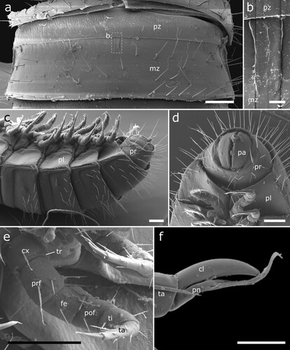
Figure 7. Siphonethus dudleycookeorum sp. nov., male holotype (ZFMK-MYR11376) from Great Britain, male sexual characters, SEM images. (a) Leg pair 2 with penes. (b) Gonopods, ventral view. (c) Gonopods, lateral view. (d) Gonopods posterior view. (e & f) detail of anterior gonopod apical podomeres. Scale: a = 20 µm, b-d = 100 µm, e = 20, f = 10 µm. Abbreviations: aI-aVI = podomeres of anterior gonopod, cx = coxa, pe = penis, pI-pVI = podomeres of posterior gonopod, prf = prefemur, st = sternite.

Figure 8. Siphonethus aff. dudleycookeorum sp. nov. (NZAC03038958), male, from New Zealand, SEM images. (a) Anterior body, ventral view. (b) Head, frontal view. (c) Detail of labrum. (d) Mid body-ring, dorsal view. (e) Posterior body-rings and preanal ring, ventral view (f) Leg pair 2 with penes, ventral view. (g) Gonopods, ventral view. Scale: a, b, d, e = 100 µm, c = 10 µm f, g = 20 µm. Abbreviations: aI-aVI = podomeres of anterior gonopod, at = antennae, co = collum, cx = coxa, gn = gnathochilarium, in = incision of labrum, la = labrum, ms = macrosetae, mz = metazonite, om = ommatidia, oz = ozopore, pa = paraproct, pe = penis, pI-pVI = podomeres of posterior gonopod, pl = pleurite, pr = preanal ring, pz = prozonite.
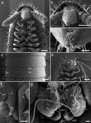
Figure 9. Siphonethus aff. dudleycookeorum sp. nov. (NZAC03038958), female, from New Zealand, SEM images. (a) Anterior body, ventral view. (b) Coxa of leg 2 with gonopore. Scale: a = 100 µm, b = 20 µm. Abbreviations: at = antennae, co = collum, cx = coxa, gp = gonopore.
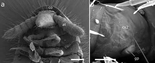
Figure 10. Head of Siphonethus coxaespinosus sp. nov. (a-c) NZAC03038955. (d & e) NZAC03038954.
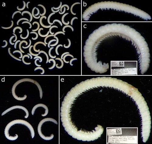
Figure 11. Siphonethus coxaespinosus sp. nov. (NZAC03038955) from New Zealand, male, SEM images. (a) Anterior body, ventral view. (b) Head, frontal view. (c) Detail of labrum. (d) Gnathochilarium, ventral view. (e) Antennae. Scale: a = 100 µm, b, c, e = 20 µm, d = 10 µm. Abbreviations: I-VII = antennomere 1-7, ac = apical cones, at = antennae; co = collum, gn = gnathochilarium, in = incision of labrum, la = labrum, ms = macrosetae, om = ommatidia sb = sensilla basiconica.
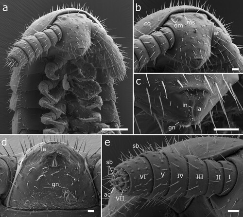
Figure 12. Body-rings and legs of Siphonethus coxaespinosus sp. nov. (NZAC03038955) from New Zealand, male, SEM images. (a) Body-ring, dorsal view. (b) Details (mesal rectangular structure) of (a). (c) Posterior body-rings and preanal ring, ventral view. (d) Anterior leg. (e) Posterior leg. Scale: a, c = 100 µm, b = 2 µm, d, e = 20 µm. Abbreviations: cl = claw, cs = coxal sacs, cx = coxa, fe = femur, mz = metauonite, oz = ozopore, pa = paraproct, pof = postfemur, pl = pleurite, pn = paronychium, pr = preanal ring, prf = prefemur, ta = tarsus, ti = tibia, tr = trochanter.
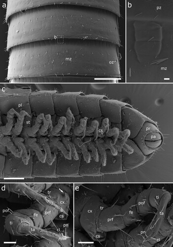
Figure 13. Male (a-e) and female (f) sexual characters of Siphonethus coxaespinosus sp. nov. from New Zealand, male, SEM images. (a) Anterior coxae of male (NZAC03038954), ventral view. (b-f) Gonopods of NAZAC03038955. (b) Ventral view. (c) Posterior view. (d) Lateral view. (e & f) Detail of apical podomere of anterior gonopod. (e) Ventral view. (f) Gonopore and 2nd leg pair of female, ventral view. Scale: a, c = 100 µm, b, d-f = 20 µm. Abbreviations: aI-aV = podomeres of anterior gonopod, bs = brush-like setae, cx = coxa, go = gonopore, pe = penis, pI-pVI = podomeres of posterior gonopod, prf = prefemur, st = sternite, stp = sternal projection, ve = external valve of vulva, vi = internal valve of vulva.
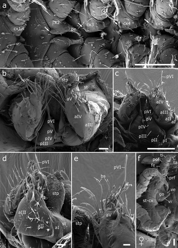
Figure 14. Siphonethus obtusus sp. nov. (NZAC03038954) from New Zealand. (a) Three male specimens). (b) Male, habitus, lateral.
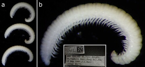
Figure 15. Head of Siphonethus obtusus sp. nov. (NZAC03038954) from New Zealand, male, SEM images. (a) Anterior body, ventral view. (b) Head, frontal view. (c) Detail of labrum. (d) Gnathochilarium, ventral view. (e) Antennae. Scale: a = 200 µm, b, c, e = 100 µm, d = 20 µm. Abbreviations: I-VII = antennomere 1-7, ac = apical cones, at = antennae; co = collum, gn = gnathochilarium, in = incision of labrum, la = labrum, ms = macrosetae, om = ommatidia, sb = sensilla basiconica.
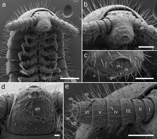
Figure 16. Body-rings and legs of Siphonethus obtusus sp. nov. (NZAC03038954) from New Zealand, male, SEM images. (a) Body-ring, dorsal view. (b) Details (mesal rectangular structure) of (a). (c) Posterior body-rings and preanal ring, ventral view. (d) Posterior leg. Scale: a, c = 100 µm, b = 10 µm, d = 20 µm. Abbreviations: cl = claw, cs = coxal sacs, cx = coxa, fe = femur, mz = metauonite, oz = ozopore, pa = paraproct, pof = postfemur, pl = pleurite, pn = paronychium, pr = preanal ring, prf = prefemur, ta = tarsus, ti = tibia, tr = trochanter.
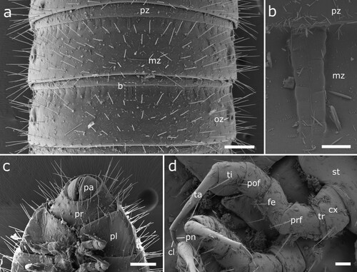
Figure 17. Male (a-d) and female (e) sexual characters of Siphonethus obtusus sp. nov. (NZAC03038954) from New Zealand, male, SEM images. (a) Coxa of leg pair 2 with gonopore, ventral view. (b-d) Gonopods. (b) Ventral view. (c) Posterior view. (d) Lateral view. (e) Female, second leg pair with gonopore, ventral view. Scale: a, e = 20 µm, b-d = 100 µm. Abbreviations: aI-aV = podomeres of anterior gonopod, cx = coxa, fe = femur, gp = gonopore, pe = penis, pI-pVI = podomeres of posterior gonopod, pof = postfemur, prf = prefemur, st = sternite, ta = tarsus, ti = tibia.
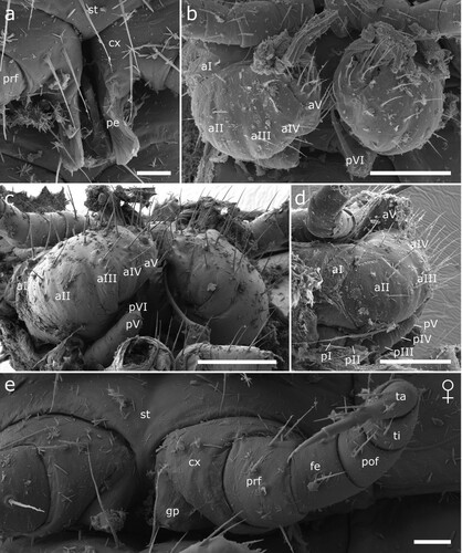
Figure 18. Siphonethus sp., specimens sequenced by Drummond et al (Citation2015). (a-c) Female (NZAC03011232). (a) Habitus, lateral. (b) Details of head. (c) Label. (d & e) Juvenile (NZAC03012795). (d) Habitus, dorsal. (e) Label.
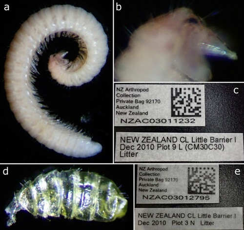
Figure 19. Siphonethus sp. (a & b) NZAC03038956. (a) Habitus, lateral. (b) Label. (c & d) NZAC3038957. (c) Habitus, lateral. (d) Label.
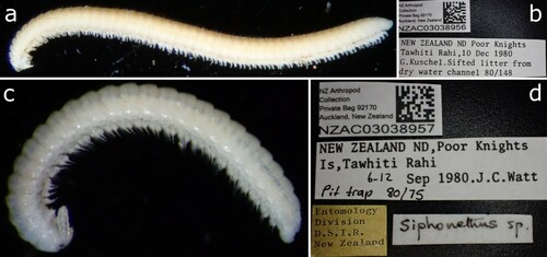
Figure 20. Maximum likelihood tree of Polyzoniida based on the COI sequence after 1000 bootstrap replicates analyzed with the Hasegawa Kinshino Yano Model. Numbers on branches indicate bootstrap support. Codon positions included were 1st+2nd+3rd. All positions with less than 5% site coverage were eliminated. The tree is drawn to scale, with branch length indicating genetic distance. Specimens from New Zealand in green box.
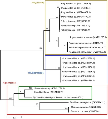
Table 3. Distance matrix. Numbers in parenthesis indicate the GenBank accession number and the voucher number.