Figures & data
Figure 1. Phylogenetic analysis of human and animal coronaviruses including the SARS-CoV-2 isolated from tiger, mink and human beings. Reproduced from Salajegheh Tazerji et al. (2020) under Creative Commons Attribution (CC BY) licence.
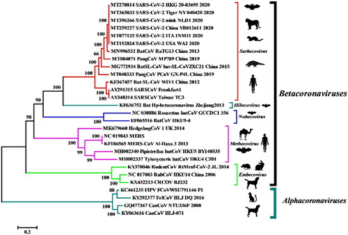
Figure 2. Countries that have reported SARS-CoV-2 infection in farmed minks. (Events in animals, Data retrieved from: https://www.oie.int/en/scientific-expertise/specific-information-and-recommendations/questions-and-answers-on-2019novel-coronavirus/events-in-animals/).
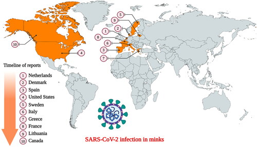
Figure 3. SARS-CoV-2 infection in mink. (A) Macroscopic image of the lungs from a mink infected with SARS-CoV-2. (B) Photomicrograph of a histopathological lung section showing interstitial pneumonia (haematoxylin and eosin, objective 20×). Reproduced from Oreshkova et al. (Citation2020) under Creative Commons Attribution (CC BY) licence.
Figure 4. Gross lung findings observed during the postmortem examination of COVID‐19 human patient. (A) Appearance of lungs in a patient with COVID‐19. The thickened alveolar septae and congestive interstitial aspects are also visible in addition to the interstitial congestion (insert). (B) Severe and extensive suppurative bronchopneumonic infiltrates (insert) observed in a patient suffering from COVID‐19. Reproduced with modifications from Menter et al. (Citation2020) under Creative Commons Attribution (CC BY) licence.
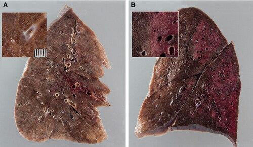
Figure 5. Electron microscopy findings. (A) Transmission electron micrograph of kidney section showing virus-like particles (sizes between 70 nm and 110 nm) within podocyte cytoplasm (arrow). (B) The vesicles contain virus‐like particles with a ring of electron‐dense granules. (C) Virus‐like particles (arrow and insert) within a vesicle that is close to the luminal border. (D) Cytoplasm of a proximal tubular epithelial cell with vesicle containing virus‐like particles (arrow and insert). Reproduced from Menter et al. (Citation2020) under Creative Commons Attribution (CC BY) licence.
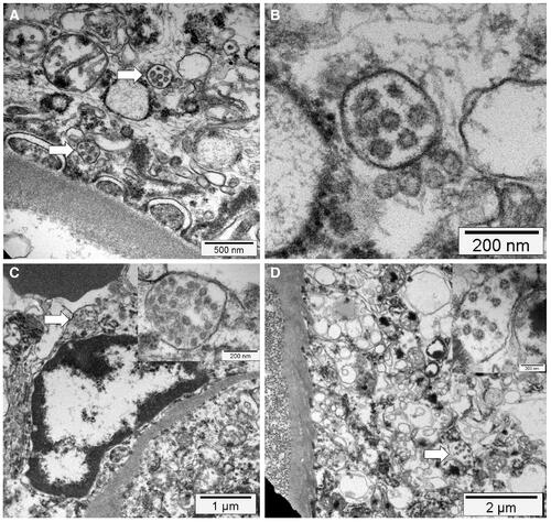
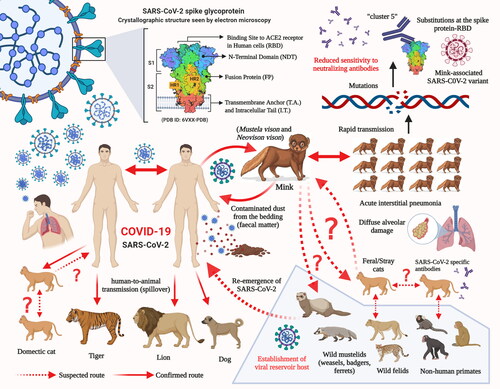
Figure 6. Potential and established routes of SARS-CoV-2 transmission between humans and animal species.