
Figures & data
Figure 1. Computed tomography of a dog diagnosed with urethral transitional cell carcinoma. (A) Sagittal view revealing thickening of the urethral wall (arrow) and enlargement of the sublumbar lymph nodes (dashed circles). Note that the enlarged lymph nodes are irregularly shaped and have heterogeneous parenchyma. (B) Plain and (C) postcontrast transverse views of the pelvic region showing an irregular and thickened urethral wall (arrows) with a hypoattenuated center and a hyperattenuated peripheral enhancement.
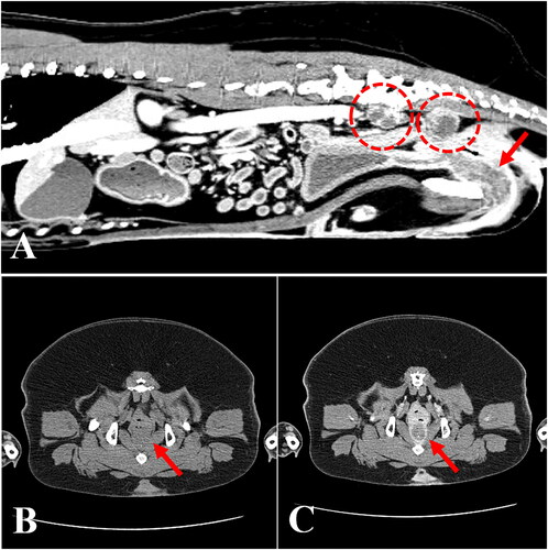
Figure 2. Gross morphological and histopathological features of the urinary bladder and proximal urethra in a dog with urethral transitional cell carcinoma. (A) The bladder mucosal wall was largely normal; however, a urethral multilobulated mass extending to the trigone area was identified (middle upper subset box). Bilateral enlarged sublumbar lymph nodes were excised in toto (middle lower picture). (B) Histopathology of the urethral mass revealed that it was composed of poorly demarcated, sessile, moderately to densely cellular, multifocally infiltrative neoplastic cells. Tumor cells were polygonal with a moderate amount of homogenous eosinophilic cytoplasm and distinct cell borders; occasionally, cells swollen due to the presence of discrete, large, clear, or eosinophilic vacuoles were observed (arrows). H&E; magnification 400×; scale bar = 50 µm. H&E, hematoxylin & eosin.
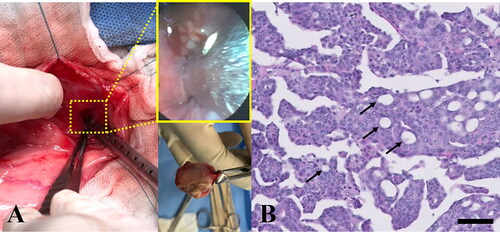
Figure 3. Canine B-RAF V595E mutation (human B-RAF V600E) in tumor and metastatic tissues. Electropherogram of samples from (A) normal tissue, (B) tumor tissue, and (C) metastatic regional lymph node tissue isolated from a dog with metastatic urethral transitional cell carcinoma at the first presentation day. c.1784T > A mutation (V595E) was detected in tumor and lymph node tissues (boxed), whereas the GTG wildtype sequence (no V595E mutation) was detected in normal tissue.
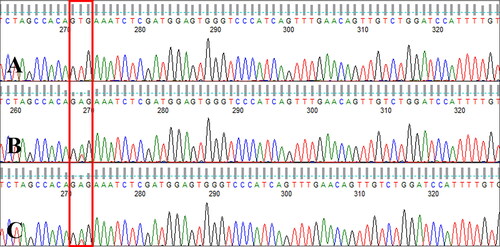
Figure 4. Canine B-RAF mutation status in ctDNA. Electropherogram of circulating cell-free DNA from serial blood samples collected during therapy with a RAF inhibitor in a dog with metastatic urethral transitional cell carcinoma at one week after the initiation of sorafenib therapy (A, week 7), during a gradual sorafenib dose escalation (B, week 11; C, week 12), and 4 weeks after the sorafenib dose was increased to 10 mg/kg BW/day (D, week 16; E, week 17). Note that the GTG wildtype sequence (no V595E mutation) was detected at week 7 and the mutant type (V595E c.1784T > A) was detected at weeks 11 and 12, whereas 4 weeks after the sorafenib dose was increased to 10 mg/kg BW/day (weeks 16 and 17), the GTG wildtype sequence was detected again (boxed).
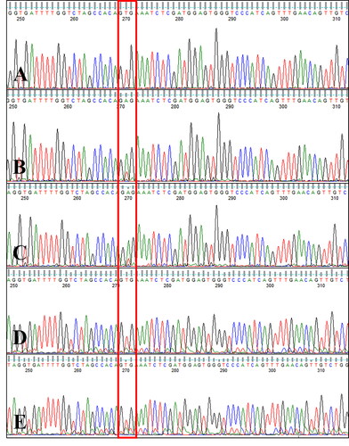
Figure 5. mRNA expression of various receptor tyrosine kinases in normal and tumor tissue samples from a dog with metastatic urethral transitional cell carcinoma. VEGFR was overexpressed in tumor compared to normal tissues. GAPDH was used for normalization. VEGFR, vascular endothelial growth factor receptor; PDGFR, platelet-derived growth factor receptors; ALK, anaplastic lymphoma kinase; EGFR, epidermal growth factor receptor.
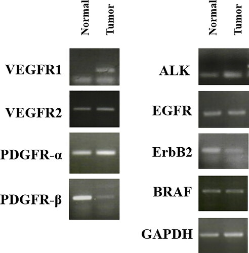
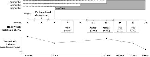
Figure 6. Changes in the dose of sorafenib administered and in B-RAF V595E-mutant circulating cell-free tumor DNA status according to the patient’s clinical course. Sorafenib was administered starting from a dose of 4 mg/kg BW/day, and the dose was gradually escalated over a period of 6 weeks. However, when dysuria worsened due to increased urethral wall thickness (asterisks) at 12 weeks postoperatively, sorafenib was escalated to the maximum tolerated oral dose of 10 mg/kg BW/day. Thereafter, the dysuria improved and the urethral wall thickness gradually decreased. The mutant (c.1784T > A) sequencing peak increased during weeks 11 and 12, concomitant with the worsened dysuria, although the mutation was not detected earlier (i.e. at 7 weeks after surgery). At 4 weeks after dose escalation to 10 mg/kg BW/day, the mutant sequencing peak was hardly detected and the wildtype (GTG) sequence was dominantly detected, concomitant with a significant decrease in urethral wall thickness and the resolution of dysuria.
Data availability statement
The data that support the findings of this study are available from the corresponding author upon reasonable request.