
Figures & data
Figure 1. The benchmark pelvic applicator consists of 12 half-wavelength dipole antennas distributed over two rings. The benchmark patient models Clarice and Will include tumors in the pelvic region. Note that the hyperthermia target volume (HTV) is the same as the gross tumor volume (GTV). The GTV is shown on the right in 3 D together with just the skeleton for easier visualization of the target.
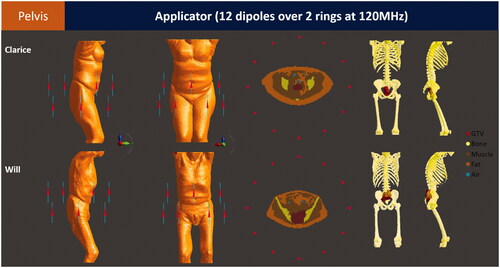
Figure 2. The benchmark H&N applicator consists of twelve half-wavelength dipole antennas distributed over three rings. The benchmark patient models Alex and Murphy include a tumor in the nasopharynx and oropharynx regions (postoperative case), respectively. Only bones and tumors are shown for clarity, please refer to and for the complete list of tissues used in the model.
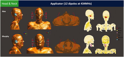
Figure 3. The benchmark breast applicator consists of 12 half-wavelength dipole antennas distributed over two rings. The benchmark patient models Venus and Luna include a superficial and a deep-seated tumor, respectively.
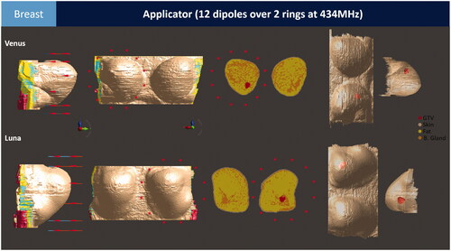
Table 1. Description of the reference and correspondent simplified benchmark applicators.
Table 2. Dielectric properties of healthy [Citation28] and tumor [Citation50] tissues at 120 and 434 MHz.
Table 3. Thermal properties of healthy and tumor tissues at baseline [Citation28] and under thermal stress [Citation56]. Blood perfusion rates are presented in SI units and in ml/min/kg for convenience.
Figure 4. THQ (Murphy, Clarice, Venus) or T50 (Alex, Will, Luna) optimized SAR and temperature distributions for the benchmark applicators displayed on top of the CT images with the target region indicated by the green contour. Axial and sagittal cross-sections are given for Murphy and Alex and axial and coronal cross-sections for Clarice, Venus, Will and Luna. Note that the CT slice of Claris and Will also includes the slings (lines, dots) on which patients are scanned and treated. The FUS results for Venus show the SAR and temperature distributions for a single pressure focus, i.e., without focus scanning.
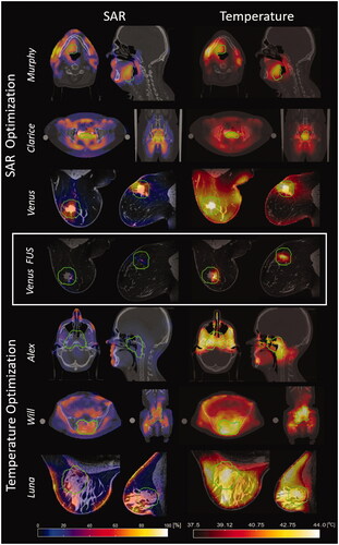
Table 4. Treatment planning results achieved using the benchmark applicators and optimized based on SAR (THQ) with VEDO software or based on temperature (tumor goal of 43 °C, with normal tissue constraints of 44 °C) with Plan2Heat software.
Table A1. Center point of the patient models, water bolus and antennas. All values are given in millimeters.