Figures & data
FIGURE 1. Tinosaurus europeocaenus, the holotypic left dentary IRSNB R 202 in A, lateral; B, medial; C, ventromedial; and D, dorsal views.
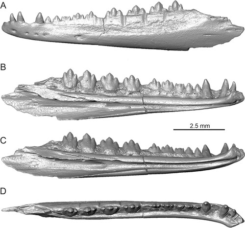
FIGURE 2. Tinosaurus europeocaenus. A–F, right maxilla IRSNB R 457 in A, lateral view; B, medial view with detail of teeth; C, ventral view; D, dorsal view; E, anteromedial view; and F, dorsoposteromedial view. G–J, left maxilla IRSNB R 458 in G, lateral; H, medial; I, ventral; and J, dorsal views.
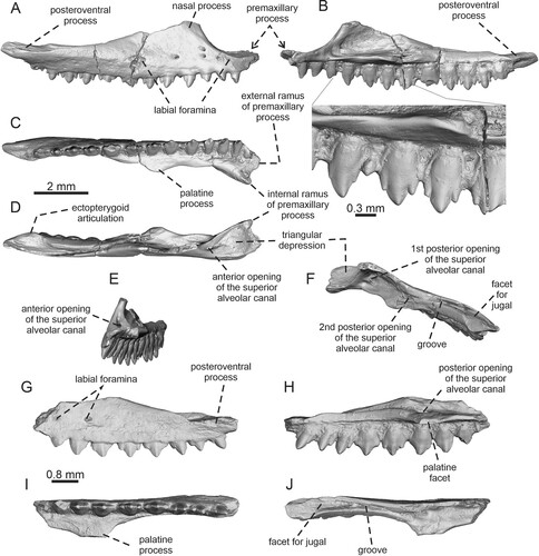
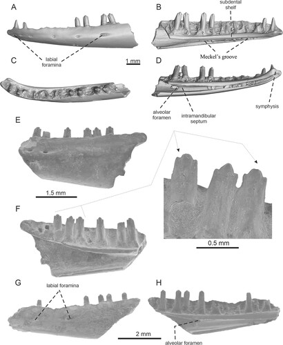
FIGURE 3. Tinosaurus europeocaenus. A–D, left dentary IRSNB R 459 in A, lateral; B, medial; C, dorsal; D, ventromedial views. E–F, right dentary IRSNB R 460 in E, lateral; and F, medial views. G–H, left dentary IRSNB R 461 in G, lateral; and H, medial views with detail of teeth.
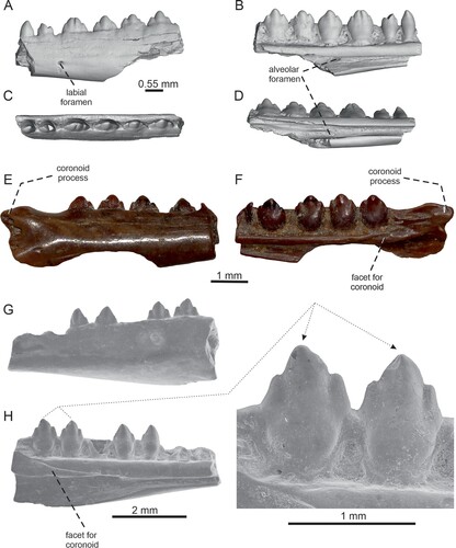
FIGURE 4. Tinosaurus europeocaenus, left dentary IRSNB R 462 showing a bony callus in A, lateral; B, medial with a detail of the callus; and C, dorsal views.

FIGURE 5. Bifurcodentodon ragei gen. et sp. nov., the holotypic left maxilla IRSNB R 463. A, lateral view with detail of teeth; B, medial view with detail of teeth; C, dorsal view; D, ventral view; E, dorsoposteromedial view with the detail of foramina set in a gutter; F, anteromedial view.
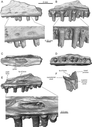
FIGURE 6. Geiseltaliellus sp., premaxilla IRSNB R 294 in A, anterior; B, posterior; and C, dorsal views.
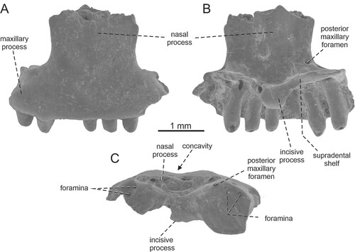
FIGURE 7. Geiseltaliellus sp. A–D, left dentary IRSNB R 464 in A, lateral; B, medial; C, dorsal; D, ventromedial views. E–F, left dentary IRSNB R 465. E, lateral view; F, medial view with detail of teeth. G–H, left dentary IRSNB R 466 in G, lateral and H, medial views.