Figures & data
FIG. 1 Fluorescence fingerprints of the tape substrate used. The fluorescence signal is divided into 15 equally spaced contour lines. The maximum fluorescence is shaded darkest. The emission is measured from 50 nm longer than the excitation wavelength to 150 nm longer than the excitation wavelength. Excitation wavelengths used are 280, 310, 340, 370, and 400 nm.
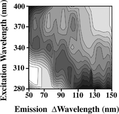
FIG. 2 Fluorescence fingerprints of the (a) dust A; (b) BC and dust A; (c) BG and dust A; (d) BP and dust A. The fluorescence signal is divided into 15 equally spaced contour lines. The maximum fluorescence is shaded darkest. The emission is measured from 50 nm longer than the excitation wavelength to 150 nm longer than the excitation wavelength. Excitation wavelengths used are 280, 310, 340, 370, and 400 nm.
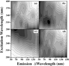
FIG. 3 Fluorescence fingerprints of the (a) Merck BG; (b) corn smut fungal spores; (c) ovalbumin. The fluorescence signal is divided into 15 equally spaced contour lines. The maximum fluorescence is shaded darkest. The emission is measured from 50 nm longer than the excitation wavelength to 150 nm longer than the excitation wavelength. Excitation wavelengths are 280, 310, 340, 370, and 400 nm.
FIG. 4 (a) PCA plot of three samples of BG (open squares), BC (open circles), BP (crosses) (9 measurements). Each sample is mixed with dust as explained in the text. Principal component 1 versus component 2 is plotted. Plots of the PCs of same three samples of BG, BC, BP (9 measurements) with the added sample of (b) Merck BG (solid diamond); (c) corn smut (solid diamond); and (d) ovalbumin (solid diamond).
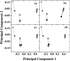
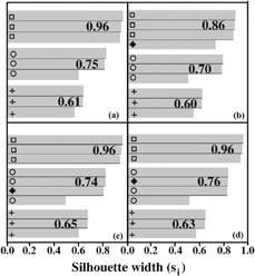
FIG. 5 (a) Silhouette plot of the strength of clustering using PAM with three samples of BG (open squares), BC (open circles), BP (crosses) (9 measurements) as shown in . Silhouette plot of the strength of clustering using PAM with the same three samples of BG, BC, BP (9 measurements) with the added sample of (b) Merck BG (solid diamond); (c) corn smut (solid diamond); and (d) ovalbumin (solid diamond). The cluster strength (Si) is explained in the text.