Figures & data
TABLE 1 Subject demographics and response to methacholine challenge
TABLE 2 Individual airway surface area, πLd, and volume factor, Ld 2 (mean ± standard error), as a function of airway generation. Measurements from the HRCT scan taken just prior to the methacholine challenge
FIG. 1 Extracted image of lung airways. This image is from a 27-year-old, healthy, nonsmoking female (height = 154 cm, weight = 59.3 kg). This subject was responsive to the methacholine challenge—with the greatest change in airway patency seen in airway generations 4, 5, and 6.
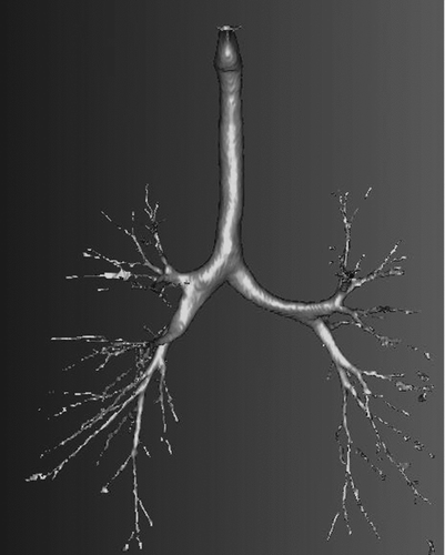
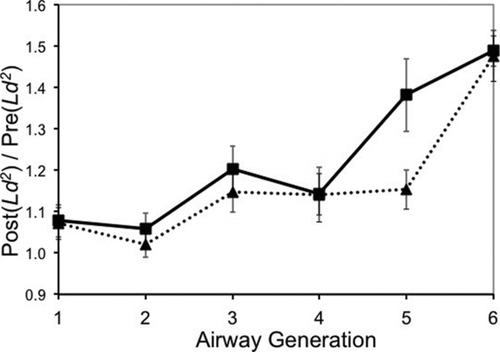
FIG. 2 Impact of bronchodilation. For each subject, morphometric measurements were made from two HRCT scans. The second HRCT scan (the “Post” scan) was made after the methacholine challenge and following bronchodilation. The first HRCT scan (the “Pre” scan) was made prior to the methacholine challenge. Post/pre airway volume factors (Ld 2; mean ± standard error) are compared: nonresponsive (6 subjects, triangles connected with dotted line); responsive (8 subjects, squares connected with solid lines).