
Figures & data
Table 1. Simulation scenarios used when calculating the charge distributions. For Scenarios 1–7 and 12–13, the flux coefficients were calculated for 35 values of ion mobility, Zi, that covered the range 0.5–2.5 cm2 V−1 s−1. For Scenarios 8 and 9, the ranges were 0.65–3.1 cm2 V−1 s−1 and 1.7–7.0 cm2 V−1 s−1, respectively. For Scenarios 10 and 11, only the mean and median, respectively, of negative and positive ion mobilities were used. For each scenario, the calculations were performed using the measured ion mobility distribution and the SR distribution, with the ion mass and mobility values corresponding to the latter given in parenthesis.
Figure 1. Processes involved in a typical particle size distribution measurement (left), and in the present simulation approach (middle); schematic illustrations of these steps (right). Note that particles with the same size but different charge state fall into different mobility sections in the signal grid, since Zp(q) = qZp(q = 1) (two topmost panels on the right). Furthermore, negative and positive particles provide separate signals, with neutral particles not detected.
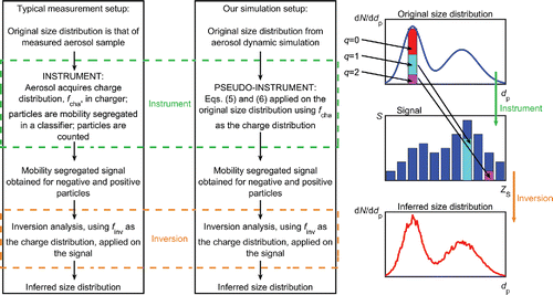
Figure 2. Top row: Time evolution of the particle size distribution in Hyytiälä (left), Mukteshwar (middle), and New Delhi (right) simulations. The color denotes the particle size distribution, dN/dlogdp, in cm−3. Bottom left: Time evolution of the particle size distribution inferred from the signal of the Hyytiälä simulation. Bottom middle: The ratio, R(dp,t), of the inverted (lower left panel) and simulated (upper left panel) distributions. The subscript “sim” and “inv” refer to the simulated and inverted distributions, respectively. Bottom right: The time averaged ratio of the inverted and simulated distributions, R*,−, and its running average, R*,−ave, according to Equations (Equation8[8] ) and (Equation9
[9] ), respectively. The data in the lower panels are shown for Hyytiälä simulation with fcha = finv and when negative particles were counted; the results for positive polarity were very similar.
![Figure 2. Top row: Time evolution of the particle size distribution in Hyytiälä (left), Mukteshwar (middle), and New Delhi (right) simulations. The color denotes the particle size distribution, dN/dlogdp, in cm−3. Bottom left: Time evolution of the particle size distribution inferred from the signal of the Hyytiälä simulation. Bottom middle: The ratio, R(dp,t), of the inverted (lower left panel) and simulated (upper left panel) distributions. The subscript “sim” and “inv” refer to the simulated and inverted distributions, respectively. Bottom right: The time averaged ratio of the inverted and simulated distributions, R*,−, and its running average, R*,−ave, according to Equations (Equation8[8] ) and (Equation9[9] ), respectively. The data in the lower panels are shown for Hyytiälä simulation with fcha = finv and when negative particles were counted; the results for positive polarity were very similar.](/cms/asset/0870d92f-13f6-40f3-80d2-574baa18e7d8/uast_a_1341039_f0002_oc.gif)
Table 2. Biases observed in the particle size distributions inferred from measurements in which the charge distribution used in the pseudo-instrument differed from that in the inversion analysis, i.e., fcha ≠ finv. The ranges of biases are given separately for negative and positive particles, R*,−ave and R*,+ave, respectively, and for particles smaller than or larger than 10 nm in diameter. The values in parenthesis were obtained using the SR distribution instead of the measured distribution. A value of R*,±ave that differs from unity reveals underestimation or overestimation of the particle concentration. The first row provides the baseline-case, i.e., fcha = finv (according to Scenario 1). In the second row, fcha was that from Scenario 1, but FHFW charge distribution was employed in the inversion. In the third row, fcha was calculated using the measured distribution, while finv was calculated using SR distribution, with both being according to Scenario 1. In other rows, the parameter that was changed when calculating fcha and finv is given in the first column, with the values used when calculating fcha and finv given in the second and third column, respectively, the other inputs were the same.
Figure 3. Left panel: The bias observed in the inferred particle size distribution when fcha was calculated using input values based on our measurements (Scenario 1 in ) and finv was according to FHFW charge distribution. The line style denotes the simulation, and signal polarity is labeled. Right panel: The same as the left panel, except that the SR distribution, instead of the measured distribution, was used when calculating fcha.
Figure 4. The bias observed in the inferred particle size distribution when fcha was calculated using the measured ion mobility distribution and finv was calculated using the SR distribution, with other inputs being the same. The line style denotes the simulation, and signal polarity is labeled.
Figure 5. Upper panels: The bias observed in the inferred particle size distribution when fcha corresponded to T and p representing conditions in laboratory (T = 298.15 K and p = 96,757 Pa; “Lab.”), at 3 km altitude (T = 269 K and p = 70,120 Pa; “3 km”), or at 10 km altitude (T = 223 K and p = 26,500 Pa; “10 km”), and finv corresponded to T and p in laboratory. The line style denotes the conditions, with markers denoting different simulations, shown only in the “10 km” case. Results corresponding to negative (positive) particle measurements are shown on the left (right). The lines depicting the “10 km” cases are practically coincident for dp < 20 nm and dp > 500 nm. Lower panels: The same as the upper panels, except that the SR distribution, instead of the measured distribution, was used when calculating fcha.
Figure 6. Left panel: The bias observed in the inferred particle size distribution when the ion masses ranged from 130 to 1000 Da (ρi = 800 kg m−3), from 220 to 1800 Da (ρi = 1500 kg m−3), or from 300 to 2700 Da (ρi = 2200 kg m−3) when calculating fcha, but were from 220 to 1800 Da when calculating finv, as indicated in the legend. Right panel: The same as the left panel, except that the SR distribution, instead of the measured distribution, was used when calculating fcha. The corresponding ion masses are given in the legend.
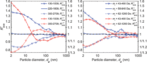
Figure 7. Left panel: The bias observed in the inferred particle size distribution when particles were assumed to be conductive, χp = ∞, or dielectric with χp = 6 or χp = 2.6 when calculating fcha, but they were assumed to be conductive when calculating finv, as indicated in the legend. Right panel: The same as the left panel, except that the SR distribution, instead of the measured distribution, was used when calculating fcha.
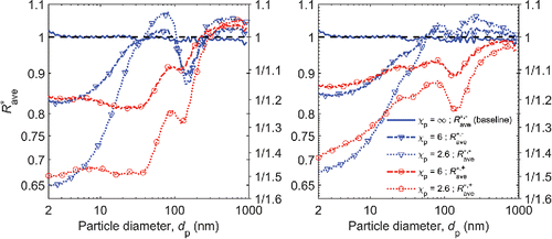
Figure 8. Left panel: The bias observed in the inferred particle size distribution when ni,T+/ni,T− was 1.0, 1.1, or 1.2 when calculating fcha, but ni,T+/ni,T− = 1.0 was used when calculating finv. The line style denotes the ni,T+/ni,T− when calculating fcha, and the marker and line color denote the signal polarity. Right panel: The same as the left panel, except that the SR distribution, instead of the measured distribution, was used when calculating fcha.

Table 3. The biases, ninv/nsim, observed in the total particle concentrations that result from using different charge distributions in the pseudo-instrument than in the inversion analysis, i.e., fcha ≠ finv. The biases are given as a range from minimum to maximum bias during the simulation followed by the bias observed at the instance of the highest particle concentration. The values in parenthesis were obtained using the SR distribution in place of the measured ion mobility distribution presented in this study. The biases are given separately for particle size ranges of 2–1000 nm and 10–800 nm, and also for particle size distributions based on counting negative and positive particles.