
Figures & data
Figure 1. A schematic illustration of the vaginal wall in healthy women (A), and cervical cancer survivors with thin epithelia and sparse dermal papillae (B). Parameters measured in each specimen; DPS: distance from dermal papilla top to epithelial surface; DPD: interdermal papilla distance; DPW: dermal papilla width; BLS: distance from basal layer to epithelial surface and the number of the dermal papillae. *p value < .05
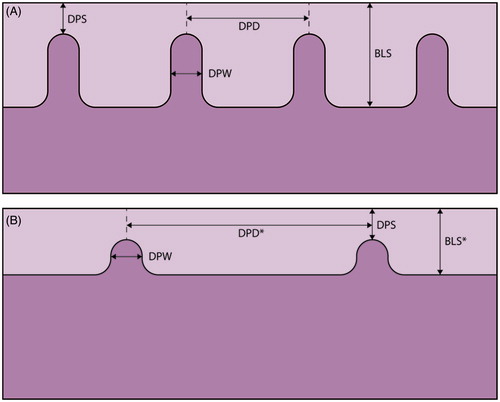
Table 1. Demographics and clinical characteristics of cervical cancer survivors and control women.
Table 2. Hormone therapy, hormonal status and clinical vaginal characteristics of cervical cancer survivors and control women.
Figure 2. Hematoxylin–eosin stained biopsies from the vaginal wall in healthy women with thick squamous cell epithelia with dermal papillae (A), and cervical cancer survivors with thin epithelia and dense connective tissue (B). Scale bar =200 μm.
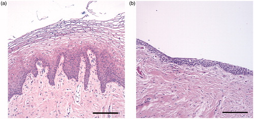
Table 3. Steroid hormone levels of cervical cancer survivors and control women.
Table 4. Sexual function, well-being, depression and anxiety in the cervical cancer survivors and the control women.