
Figures & data
Figure 1. The structure of Networks. (a) Dense Net, (b) V-Net, (c) Dense V-Network.
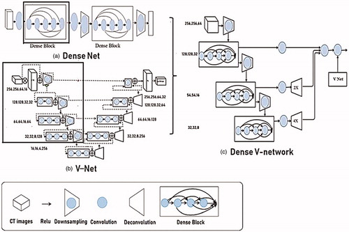
Figure 2. The model training process.
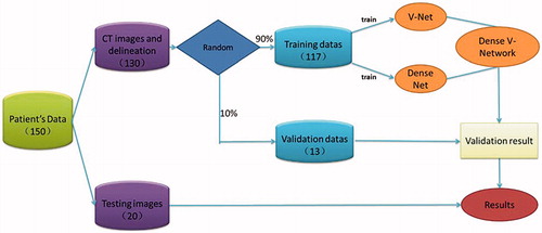
Figure 3. The box plot of three evaluation parameter results.
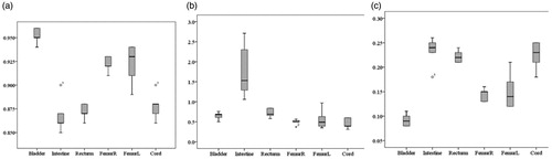
Table 1. Segmentation results of pelvic organs at risk (Mean).
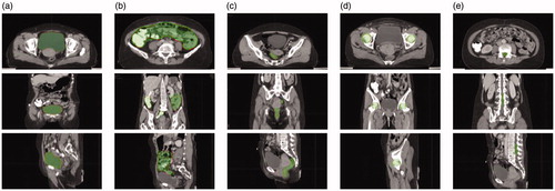
Figure 4. The automatic segmentation result of CT images. (a) Bladder, (b) intestine, (c) rectum, (d) femur, (e) cord.
Supplemental material