1,683
Views
23
CrossRef citations to date
0
Altmetric
Original Articles

Epidemiologic assessment of trauma-independent skeletal pathology in non-passerine birds from museum collections

&
Pages 212-219 | Received 01 Jan 2005, Accepted 02 Mar 2005, Published online: 19 Oct 2010

Figures & data

Table 1. Distribution of avian osseous infection

Figure 1. Posterior view of Macaroni penguin (Eudyptes chrysolophus) ankle. Reactive new bone of infectious arthritis.
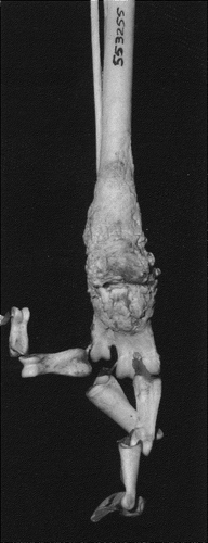
Figure 1. Posterior view of Macaroni penguin (Eudyptes chrysolophus) ankle. Reactive new bone of infectious arthritis.

Figure 2. Posterior view of emu (Dromaius novaehollandiae) knee. Severe disruptive changes of infectious arthritis.
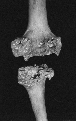
Figure 2. Posterior view of emu (Dromaius novaehollandiae) knee. Severe disruptive changes of infectious arthritis.

Figure 3. Inferior view of common rhea (Rhea Americana) femoral condyle. Focal articular surface defect from osteochondritis dessicans.
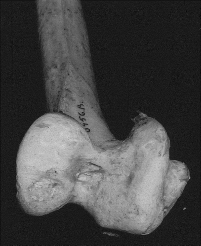
Figure 3. Inferior view of common rhea (Rhea Americana) femoral condyle. Focal articular surface defect from osteochondritis dessicans.

Figure 4. Posterior view of Bateleur eagle (Terathopius ecaudatus) metatarsus. Prominent mid diaphyseal bump, related to neoplasm.
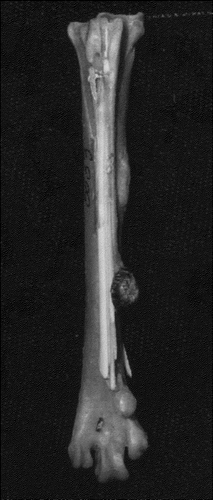
Figure 4. Posterior view of Bateleur eagle (Terathopius ecaudatus) metatarsus. Prominent mid diaphyseal bump, related to neoplasm.

Figure 5. Right lateral view of ostrich (Struthio camelus) chest. Large malignant calcific masses.

Figure 5. Right lateral view of ostrich (Struthio camelus) chest. Large malignant calcific masses.

Figure 6. Lateral view of hard head duck (Aythya australis) upper extremities. Diffuse enlargement with irregular lacy surface, from probable neoplasm.
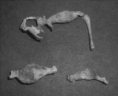
Figure 6. Lateral view of hard head duck (Aythya australis) upper extremities. Diffuse enlargement with irregular lacy surface, from probable neoplasm.

Figure 7. Lateral view of skeletonized body of bare-faced curassow (Crax fasciolata). Osteolytic areas from cancer.
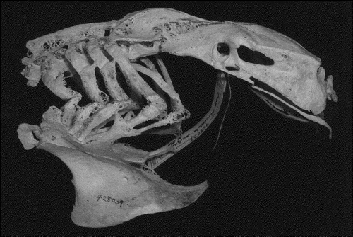
Figure 7. Lateral view of skeletonized body of bare-faced curassow (Crax fasciolata). Osteolytic areas from cancer.

Figure 8. Ventral view of snow goose (Anser cygnoides) pelvis. New bone formation of a mass.

Figure 8. Ventral view of snow goose (Anser cygnoides) pelvis. New bone formation of a mass.

Figure 9. Anterior oblique view of red-necked pheasant (Phasianus colchicus) distal tibiotarsus. Periosteal thickening with isolated spicules from neoplasm.
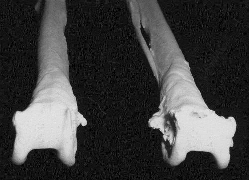
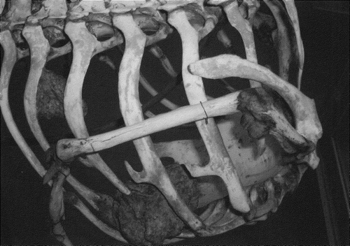
Figure 9. Anterior oblique view of red-necked pheasant (Phasianus colchicus) distal tibiotarsus. Periosteal thickening with isolated spicules from neoplasm.
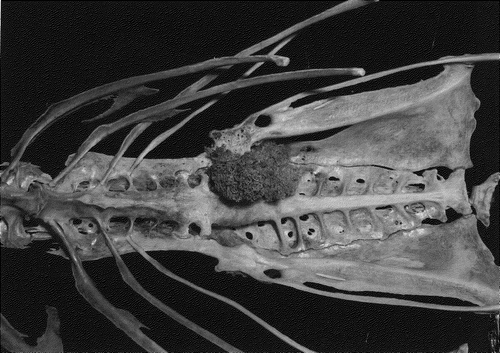
Figure 10. Anterior view of Australian cassowary (Casuarius casuarius) proximal tibiotarsus. Focal spheroid lesion with overhanging edge classic for gout.

Figure 10. Anterior view of Australian cassowary (Casuarius casuarius) proximal tibiotarsus. Focal spheroid lesion with overhanging edge classic for gout.

Figure 11. Medial oblique view of black-throated diver (Gavia arctica) distal tibiotarsus. Overgrowth at joint margin forming osteophytes of osteoarthritis.

Figure 11. Medial oblique view of black-throated diver (Gavia arctica) distal tibiotarsus. Overgrowth at joint margin forming osteophytes of osteoarthritis.

Reprints and Corporate Permissions

Please note: Selecting permissions does not provide access to the full text of the article, please see our help page How do I view content?

To request a reprint or corporate permissions for this article, please click on the relevant link below:

Academic Permissions

Please note: Selecting permissions does not provide access to the full text of the article, please see our help page How do I view content?

Obtain permissions instantly via Rightslink by clicking on the button below:

If you are unable to obtain permissions via Rightslink, please complete and submit this Permissions form. For more information, please visit our Permissions help page.