Figures & data
Table 1. Synthesis of all the advantages and strengths of using ultrasound in COVID-19.
Figure 1. Bedside evaluation by a primary care physician of a patient with SARS-CoV-2 infection: B-lines at lung ultrasound appear as slightly hyperechoic bundles perpendicular to the hyperechoic pleural line.
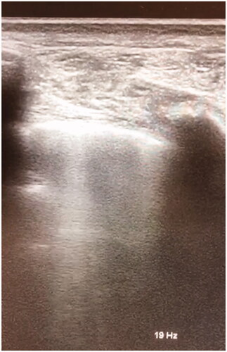
Figure 2. Pleural effusion at lung ultrasound in a SARS-CoV-2 patient appears as an anechoic area (on the right region of the picture).
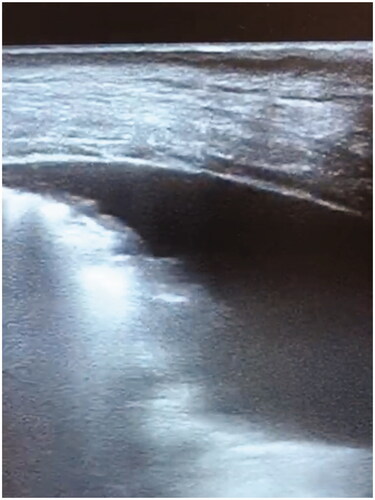
Table 2. Comparison of LUS and CT scan imaging in the assessment of COVID-19 patients.
Figure 3. The Italian network between primary care physicians and USCA doctors, who are activated by general practitioners to evaluate at home COVID-19 patients. The USCA doctors in presence of high risk factors, signs suggestive of respiratory failure and bilateral pneumonia could activate emergency system to require hospitalisation for COVID-19 patients in HUB dedicated medical centres.
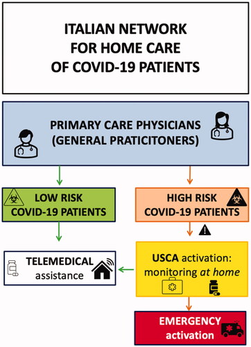
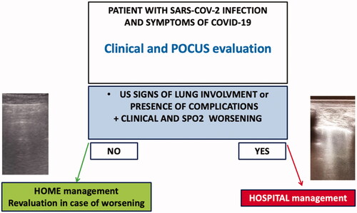
Figure 4. Management of patients with SARS-CoV-2 in primary care. The presence of ultrasonographic signs of lung involvements or the presence of complications (for example, pleural effusions and cardiovascular alterations) in addition to clinical and SpO2 worsening could suggest the physicians to in-hospital management of patients.