5,534
Views
129
CrossRef citations to date
0
Altmetric
Review Article

Rheumatoid arthritis and the complement system

, , &
Pages 517-530 | Published online: 08 Jul 2009

Figures & data

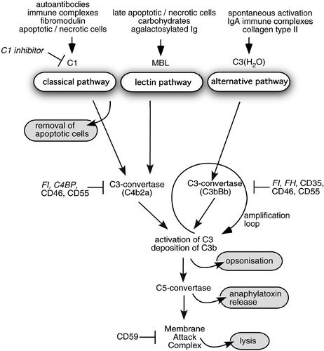
Figure 1. Scheme of the complement system and its inhibitors. Three pathways by which the human complement system can be activated and their physiological effects: 1) clearance of apoptotic cells, 2) opsonization of pathogens and immune complexes for phagocytosis, 3) release of anaphylatoxins and lysis. Furthermore, sites of action of soluble (italics) and membrane‐bound (regular font) complement inhibitors are indicated.

Figure 1. Scheme of the complement system and its inhibitors. Three pathways by which the human complement system can be activated and their physiological effects: 1) clearance of apoptotic cells, 2) opsonization of pathogens and immune complexes for phagocytosis, 3) release of anaphylatoxins and lysis. Furthermore, sites of action of soluble (italics) and membrane‐bound (regular font) complement inhibitors are indicated.

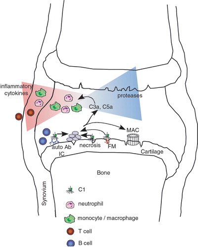
Figure 2. Schematic diagram of possible contributions of the complement system to development of rheumatoid arthritis(RA) in a joint. Complement can be activated in several ways in the RA joint. Examples are deposited autoantibodies and immune complexes (IC), apoptotic and necrotic cells, certain cartilage molecules such as fibromodulin (FM) exposed upon initial cartilage damage. Activation of complement leads to a release of proinflammatory anaphylatoxin C5a that attracts and activates neutrophils and macrophages. These cells in turn release proteases and cytokines attracting T and B lymphocytes and other inflammatory cells, further fuelling the process of inflammation. As a result of complement activation membrane attack complex (MAC) is formed and has a plethora of effects on cells even at sublytic concentrations.

Figure 2. Schematic diagram of possible contributions of the complement system to development of rheumatoid arthritis(RA) in a joint. Complement can be activated in several ways in the RA joint. Examples are deposited autoantibodies and immune complexes (IC), apoptotic and necrotic cells, certain cartilage molecules such as fibromodulin (FM) exposed upon initial cartilage damage. Activation of complement leads to a release of proinflammatory anaphylatoxin C5a that attracts and activates neutrophils and macrophages. These cells in turn release proteases and cytokines attracting T and B lymphocytes and other inflammatory cells, further fuelling the process of inflammation. As a result of complement activation membrane attack complex (MAC) is formed and has a plethora of effects on cells even at sublytic concentrations.

Table I. Evidence for involvement of complement in rheumatoid arthritis (RA) observed in animal models.

Table II. Therapeutic approaches to complement inhibition in rheumatoid arthritis.
Reprints and Corporate Permissions

Please note: Selecting permissions does not provide access to the full text of the article, please see our help page How do I view content?

To request a reprint or corporate permissions for this article, please click on the relevant link below:

Academic Permissions

Please note: Selecting permissions does not provide access to the full text of the article, please see our help page How do I view content?

Obtain permissions instantly via Rightslink by clicking on the button below:

If you are unable to obtain permissions via Rightslink, please complete and submit this Permissions form. For more information, please visit our Permissions help page.