Figures & data
Table 1. Concentrations of different adipokines measured in SGBS supernatants (IL, interleukin; TNF, tumor necrosis factor) used for stimulation, harvested on day 10 of adipogenesis. Analyses were performed using Thermo Fisher Scientific’s LPrecellys® technology.
Figure 1. Representative snapshot of control NK-92 cells without (a) and with (b) tracking mode as seen via CytoSMARTTM live cell imaging technology.
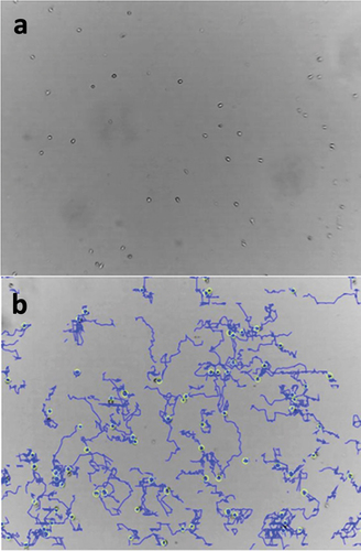
Figure 2. a) Representative Western Blot analysis of GAPDH and CFL-1 after the transfection of CFL-1 siRNA constructs into NK-92 cells using LipocalyxViromer®BLUE. Protein levels of CFL-1 (b), pCFL-1 (c) after Viromer®BLUE-mediated CFL-1 knockdown. NK-92 cells were incubated with Viromer®BLUE reagents and either CFL-1 siRNA construct #1, CFL-1 siRNA construct #2 or silencer negative control siRNA for 24 hours (negative control). NK-92 cells incubated with RPMI only were considered as controls. All data represent means and standard error of means (±SEM). Data was pooled from four independent replicates.
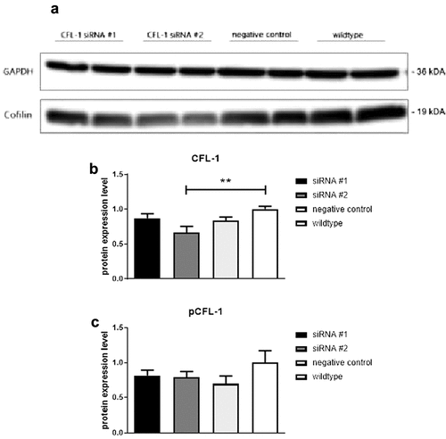
Figure 3. Accumulated distance in µm (a) and mean velocity in µm/min (b) shown by transfected NK-92 cells that exhibited significantly reduced CFL-1 expression levels. Migration activity was recorded for 60 minutes (1 picture per minute) via CytoSmart analyses in control cells and CFL-1 siRNA #2 transfected cells. All data represent means ±SEM. Data was pooled from four independent replicates.
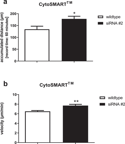
Figure 4. Protein levels of beta-actin (a), CFL-1 (b) and inactive pCFL-1 (c) as well as ratios of pCFL-1/CFL-1 (d) after short-term SGBS adipocyte-conditioned medium stimulation analyzed by Western Blot. NK-92 cells were incubated for 0.5 hours with serial dilutions of SGBS adipocyte-conditioned medium (harvested on day 10 of adipogenesis, pure or diluted 1:10, 1:100 and 1:1000 in RPMI, each with 250 µl). NK-92 cells incubated with RPMI only served as controls. All data represent means ± SEM. Data was pooled from three independent replicates.
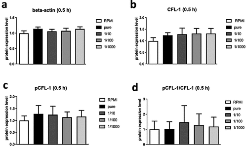
Figure 5. Accumulated distance in µm (a) and mean velocity in µm/min (b) of NK-92 cells after short-term stimulation with SGBS ACM adipocyte-conditioned medium. Adherent NK-92 cells were incubated with RPMI supplemented with 250 µl of either pure or 1:100 diluted in RPMI SGBS adipocyte-conditioned medium (harvested on day 10 of adipogenesis). Migration activity was recorded for 60 minutes (1 frame per minute) via CytoSmart analyses. NK-92 cells treated with RPMI only served as controls. All data represent means ± SEM. Data were pooled from three independent replicates.
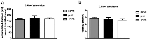
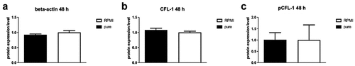
Figure 6. Protein levels of ß-actin (A), CFL-1 (B) and inactive pCFL-1(C) after long-term SGBS adipocyte-conditioned medium stimulation. NK-92 cells were incubated for 48 hours with RPMI supplemented with 250 µl pure SGBS adipocyte-conditioned medium (harvested on day 10 of adipogenesis). NK-92 cells incubated with RPMI only served as controls. All data represent means ± SEM. Data was pooled from three independent replicates.