Figures & data
Figure 1. (A) A light microscopic image from a selected liver section. (B) Light microscopic image at (A) after superimposing a point-counting grid on it. (C and D) Stereological procedures for sampling and applying the physical dissector. C reference and D are look-up sections.*Section profiles of glomeruli in the reference and look-up sections. The black arrow was counted as a dissector particle if its profile was not seen in the look-up section. The arrow with a white filling sign profile of the particle in the look-up section was not seen in the reference section.
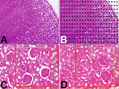
Figure 2. The volumetric values of all groups ± SEM are summarized.
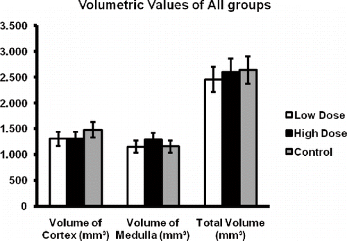
Figure 3. The mean numerical density of glomeruli ± SEM in all groups is summarized.
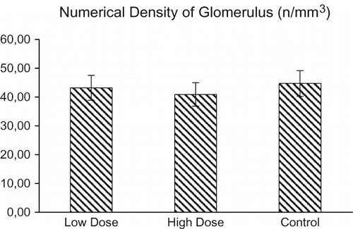
Figure 4. The total number of glomeruli ± SEM in all groups is summarized.
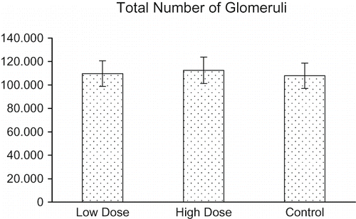
Figure 5. The mean height of glomerulus ± SEM in all groups is summarized.
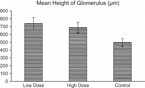
Figure 6. Light microscopy of kidneys in the control group. In this figure, glomeruli and tubuli with healthy appearance are seen. Abbreviations: g = glomerulus, d = distal tubules, p = proximal tubules, t = collecting tubules. *Blood vessel. Dye: Hematoxylin-eosin; Magnification bars: 50 μm.
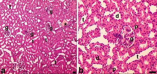
Figure 7. Light microscopy of kidney in the low-dose haloperidol-given group. Arrow heads: cells of tubules that have pyknotic nuclei and eosinophilic cytoplasm. *Hydropic vacuolation in tubule cells. Abbreviations: t = tubular lumen, boxed areas subjected to focal necrosis; d = distal tubule surrounded with vacuolated cells; n = necrotic tubules and necrotic tubule cells; arrow = desquamated cell in the lumen; e = eosinophilic accumulation in proximal tubule lumen and vacuolated tubule cells; ˆ = dilated Bowman space. Circled area = space that occurred after mesangial cells damage; + = dilated glomerular capillaries. Also in B, + indicates thickening in the basal membrane of the intraglomerular capillary. The inset shows boxed areas of A at high magnification. Dye: Hematoxylin-eosin; Magnification bars: 40 μm.
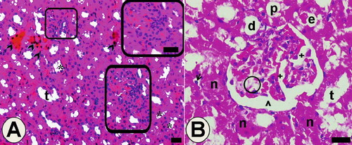
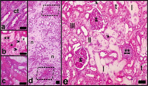
Figure 8. Light microscopy of kidney in the high-dose haloperidol-given group. Abbreviations: ct = enlarged connective tissue among the necrotic tubules. **Necrotic distal tubule. *Necrotic proximal tubule. Black arrow head = tubular basal membrane thickening and hyaline deposits; white arrows = venous thrombus and hyaline deposit-filled necrotic tubules. d indicates general view of renal medulla; n = necrotic areas, limited areas, blood vessels, and damaged tubules. e indicates a general view of the renal cortex. *Mesangial cell proliferation and focal necrosis in glomeruli; **Damaged glomerulus. t = tubules with hydropic degeneration; III, II, I = necrotic tubules at increasing degrees (the most in III). Arrows = tubular basal membrane thickening and hyaline deposits. Dye: Hematoxylin-eosin; Magnification bars: 50 μm.