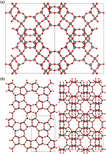
5,770
Views
62
CrossRef citations to date
0
Altmetric
Articles

DL_MONTE: a general purpose program for parallel Monte Carlo simulation

, &
Pages 1240-1252 | Received 20 May 2013, Accepted 05 Aug 2013, Published online: 25 Sep 2013

Figures & data

Figure 1 The time taken for two representative simulations. The first is for GCMC simulation of CO2 in zeolite-A (approximately 2400 zeolite ions and 100 CO2 molecules) and the second for the NPT simulation of bulk MgO (512 ions). Both calculations used the Ewald summation technique. The zeolite simulations were carried out on the UK supercomputing facility, HECToR, a Cray XE6 and the MgO simulation on the IBM Idataplex, Blue Wonder, at Daresbury Laboratory. All times are referenced to those taken on a single core.
Figure 2 The enthalpy of forming, ΔH (kJ mol–1), for different size nanoparticles. All calculations were carried out at 800 K.
Figure 3 (Colour online) Structure of 21,952 ion nanoparticle containing 5% Mn2+ ions. The Mn2+ ions preferentially decorate corners, edges and faces. Mg2+, Mg2+ and O2– are coloured green, purple and red, respectively.
Figure 4 The calculated phase diagram of the 1728 nanoparticle. The solid line is the binodal and the dashed line the spinodal.
Figure 5 The consolute temperature Tc as a function of nanoparticle size (total number of ions).
Figure 6 The average number of adsorbed water molecules, as a function of the chemical potential, . The calculation of is described in the text. All the calculations were performed at 350 K.
Figure 7 (Colour online) The distribution of water molecule energies determined at  kJ mol–1 and  kJ mol− 1. The energies displayed are taken from representative simulations at 275 K.
Figure 8 (Colour online) The structure of water molecules at the surface for (a)  = 69 and (b)  = 86. The Mg2+ and O2– ions in the substrate are coloured green and red, respectively. The O and H atoms in the water molecule are coloured blue and white, respectively.
Figure 9 (Colour online) Structure of (a) FAU viewed along (1 1 0) and (b) MFI viewed along (0 1 0) and (1 0 0). Grey colour represents silicon and red colour represents oxygen.

Table 1 Simulated zeolite lattice parameters compared to experiment.

Figure 10 (Colour online) Adsorption of CO2 in Na-FAU (a) adsorption isotherms, (b) adsorption isosteres and (c) heats of adsorption.
Figure 11 (Colour online) Adsorption isotherms of CO2 in siliceous MFI, coloured according to temperature. Data points are our simulations, line is experimental data from Yamazaki et al. [Citation54] at 273 K.

Table 2 Sensitivity on potential parameters of the number of CO2 molecules adsorbed at two test pressures, 5 and 25 bar, to compare with experimental data from Maurin et al. [Citation41].

Figure 12 (Colour online) Heat of adsorption of CO2 in Na-FAU, Si:Al ratio = 1. Green: test 13, blue: test 14, red: test 15, orange: experimental data from Maurin et al. [Citation41].

Table 3 Lennard-Jones potential parameters and partial charges.
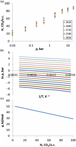
Figure 13 (Colour online) Adsorption isotherm of CO2 in Na-FAU with Si:Al ratio = 1 (green) and Si:Al ratio = 1.18 (red and blue) at 303.2 K. Data points are our simulations, blue line is represents experimental data from Pillai et al. [Citation55].