
Figures & data
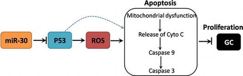
Fig. 1. Expression of miR-30 in GC tissues and cell lines.
Notes: (A) Relative expression of miR-30 in all tissue samples of GC and normal tissues. (B) Relative expression of miR-30 in GES-1, HGC-27, MKN-28, MKN-45, MGC-803, SGC-7901 cell lines. Results were shown as the mean ± SEM. *Indicated statistical significance (p < 0.05) compared with respective control.
Fig. 2. Effect of up- and down-regulation of miR-30 on cell proliferation and apoptosis in HGC-27 cells.
Notes: HGC-27 cells were transfected with miR-30 inhibitors or mimics. (A and C) Efficiency of inhibition or overexpression of miR-30 expression was examined by real-time PCR. Relative expression of miR-30 was shown as folds of control. (B and D) Cell proliferation was measured by an assay kit and cell growth curve was shown. (C and E) Apoptotic cell death was measured using TUNEL staining and results were shown as folds of control. Results were shown as the mean ± SEM. *Indicated statistical significance (p < 0.05) compared with respective control.
Fig. 3. Role of P53 in the effect of down-regulation of miR-30 on cell proliferation and apoptosis in HGC-27 cells.
Notes: (A) Relative expression of P53 in all tissue samples of GC and normal tissues. (B) Correlation of the expression of miR-30 and P53 in GC tissues. HGC-27 cells were transfected with miR-30 inhibitors. mRNA (C) and protein (D) expression of P53 was determined by real-time PCR and western blot. HGC-27 cells were transfected with shP53 lentivirus and then transfected with miR-30 inhibitors. (E) Cell proliferation was measured by an assay kit and cell growth curve was shown. (F) Apoptotic cell death was measured using TUNEL staining and results were shown as folds of control. Results were shown as the mean ± SEM. *Indicated statistical significance (p < 0.05) compared with respective control. #Indicated statistical significance (p < 0.05) compared with miR-30 inhibitor group.
Fig. 4. Role of P53/ROS-mediated mitochondrial pathway in the effect of downregulation of miR-30 on cell proliferation and apoptosis in HGC-27 cells.
Notes: HGC-27 cells were transfected with shP53 lentivirus and then transfected with miR-30 inhibitors. (A) cells were stained with 10 μM DHE to examine ROS level. HGC-27 cells were transfected with shP53 lentivirus or pretreated with100 μM NAC, and then transfected with miR-30 inhibitors. (B) Mitochondria were isolated and oxygen consumption rate was determined using a Clark Oxygen Electrode. (C) Cytoplasmic protein was extracted and protein expression of Cyto C was determined by western blot. (D and E) Caspase 3 and 9 activities were determined using commercial kits. (F) Apoptotic cell death was measured using TUNEL staining and results were shown as folds of control. Results were shown as the mean ± SEM. *Indicated statistical significance (p < 0.05) compared with respective control. #Indicated statistical significance (p < 0.05) compared with miR-30 inhibitor group.
Fig. 5. Schematic figure of the mechanism of miR-30-mediated regulation of GC cell proliferation.
Supplemental material