Figures & data
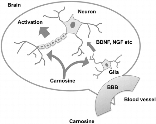
Figure 1. BBB permeability of IDP and activation of neuronal cells by carnosine. (A) BBB permeability of IDP. BBB permeability of IDP was assessed using a BBB kit and estimated by the resulting permeability coefficient (Papp). (B) Activation of SH-SY5Y cells by carnosine. Carnosine was added to SH-SY5Y cells, and neurite (arrow) growth was observed by fluorescence microscopy (BZ-X700, Keyence, Osaka, Japan).
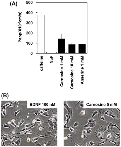
Figure 2. Carnosine augments the expression of neurotrophin genes. The effects of carnosine on expression of BDNF in U-87 MG cells (A) and SH-SY5Y cells (B) were evaluated by qRT-PCR. The effects of carnosine on the expression of NGF (C and D), NTF4 (E and F) and GDNF (G and H) in U-87 MG cells and SH-SY5Y cells, respectively, were also evaluated by qRT-PCR.
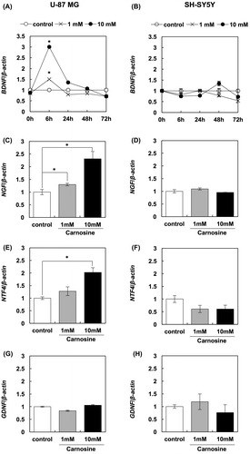
Figure 3. Carnosine activates BDNF production in U-87 MG cells. U-87 MG cells were treated with 10 mM carnosine for 3 days. BDNF in the supernatant was assessed using an ELISA kit (Promega).
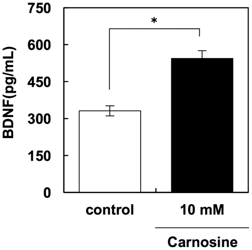
Figure 4. Carnosine promotes neurite outgrowth in neuronal cells via glial cells. After U-87 MG cells were treated with 5 mM carnosine for 24 h, supernatant was collected and added to SH-SY5Y cells. BDNF (100 ng/mL) was used as a positive control. Cells were stained with Milli-Mark FluoroPan Neuronal Marker (A). Neurite length was determined using a Neurite Outgrowth Assay kit (B).
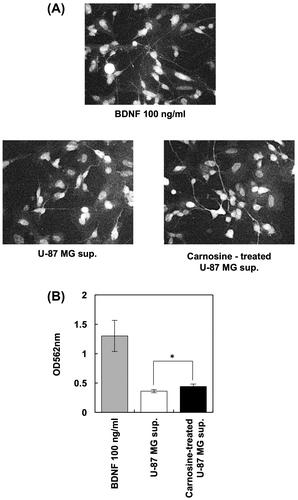
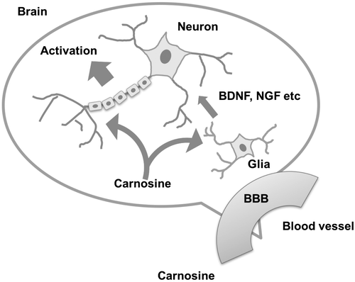
Figure 5. Schematic diagram of carnosine function in the brain.