Figures & data
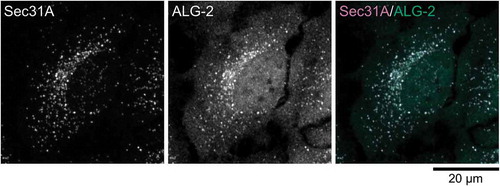
Figure 1. ERES localization of ALG-2. (a) Schematic representation of COPII vesicle formation in the ERES. (b) Representative micrograph showing the ERES localization of ALG-2. HeLa cells were fixed with 4% paraformaldehyde in phosphate buffer, permeabilized with 0.1 % Triton X-100, and then triple stained with antibodies against an ER marker, calnexin (a rabbit polyclonal antibody, Enzo Life Sciences), an ERES marker, Sec31A (a mouse monoclonal antibody, BD Biosciences) and ALG-2 (a goat polyclonal antibody) [Citation98]. The images are superimpositions of serial optical sections taken by a confocal laser-scanning microscope through the whole thickness of the cell. The right panel shows a merged image with the pseudocolors as follows: green (ALG-2) and magenta (Sec31A).
![Figure 1. ERES localization of ALG-2. (a) Schematic representation of COPII vesicle formation in the ERES. (b) Representative micrograph showing the ERES localization of ALG-2. HeLa cells were fixed with 4% paraformaldehyde in phosphate buffer, permeabilized with 0.1 % Triton X-100, and then triple stained with antibodies against an ER marker, calnexin (a rabbit polyclonal antibody, Enzo Life Sciences), an ERES marker, Sec31A (a mouse monoclonal antibody, BD Biosciences) and ALG-2 (a goat polyclonal antibody) [Citation98]. The images are superimpositions of serial optical sections taken by a confocal laser-scanning microscope through the whole thickness of the cell. The right panel shows a merged image with the pseudocolors as follows: green (ALG-2) and magenta (Sec31A).](/cms/asset/2189ad18-be30-48db-ab6f-db349c3cc347/tbbb_a_1525274_f0001_oc.jpg)
Figure 2. Structures of ALG-2. (a) Schematic representation of primary structure of ALG-2. ALG-2 has an N-terminal extension rich in Ala, Gly and Pro, and a C-terminal penta-EF-hand (PEF) domain. Calcium ions (Ca2+) bind to EF1 and EF3 under physiological conditions. ALG-2ΔGF122 is an alternatively spliced variant of ALG-2 lacking two amino acids, Gly121 and Phe122. (b) Co-crystal structure of the complex between ALG-2 and Sec31A peptide (PDB code, 3WXA). Chain A and B (ALG-2 molecule A and B) are shown by cartoon (green, EF1 and EF3; dark green, EF5, dark grey, EF2 and EF4) and surface representation (blue, pocket 1; light blue, pocket 2; green, pocket 3), respectively, using PyMOL software. Chain C and D (Sec31A peptides) are shown by stick in magenta. Grey spheres indicate zinc ions (Zn2+). (c) Schematic representation of the ALG-2 dimer-mediated physical association between two target proteins. The ALG-2 dimer couples two target proteins in a Ca2+-dependent manner.
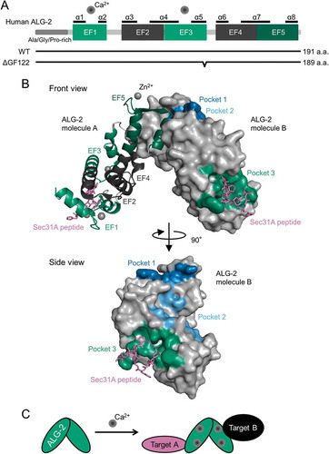
Table 1. Combinations of proteins bridged by ALG-2 homodimer and ALG-2-peflin heterodimer.
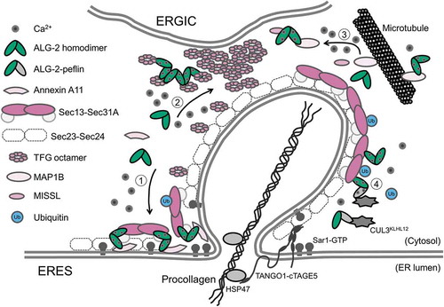
Figure 3. Schematic representation of a potential model for adaptor functions of ALG-2 in the ERES. The ALG-2 homodimer couples following combinations of target proteins: 1) Sec31A and annexin A11, 2) two TFG octamers, and 3) MISSL and MAP1B, whereas 4) the ALG-2-peflin heterodimer bridges between Sec31A and CUL3KLHL12 in a Ca2+-dependent manner.