Figures & data
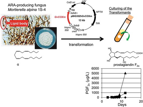
Figure 1. Schematic diagram of the reactions from ARA to PGF2α in the COX enzyme system.
PGG2, prostaglandin G2; PGH2, prostaglandin H2; PGF synthase, prostaglandin F synthase; PGF2α, prostaglandin F2α.
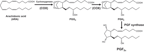
Figure 2. Schematic diagram of the plasmid pBIG35ZhGvCOXm with the GvCOX gene downstream of the histone promoter.
GvCOXm, the codon-optimized GvCOX gene for M. alpina; hispro550 p, M. alpina histone protein promoter; SdhB t, M. alpina SdhB gene transcription terminator; ura5, the orotate phosphoribosyl transferase gene of M. alpina 1S-4; NPTIII, neomysin phosphotransferase III gene; TrfA, TrfA locus, which produces two proteins that promote the replication of the plasmid; ColE1 ori, ColE1 origin of replication; oriV, pRK2 origin of replication; RB and LB, the right and left border, respectively, from Ti-plasmid of A. tumefaciens.
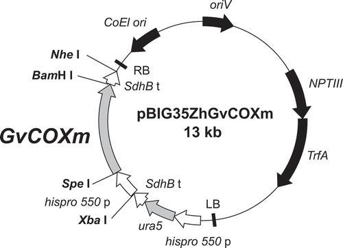
Figure 3. Evaluation of in vitro COX activity in the transformants.
Resting cells were used to convert 30.45 mg/L of ARA to PGF2α as described in Materials and Methods.
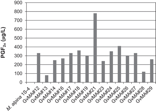
Figure 4. Evaluation of fermentative production of PGF2α that was released into the media during the 12-day cultivation of the transformants.
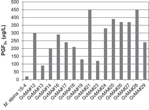
Figure 5. Fermentative intracellular production of prostaglandins F1α, F2α, and F3α in the parental (M. alpina 1S-4) and transformant (GvMA#21 and GvMA#28) strains.
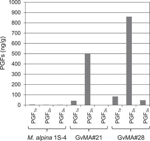
Figure 6. Fermentative extracellular production of prostaglandins F1α, F2α, and F3α in the parental (M. alpina 1S-4) and transformant (GvMA#21 and GvMA#28) strains.
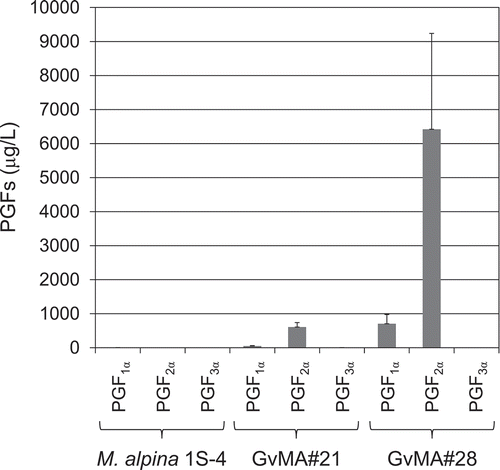
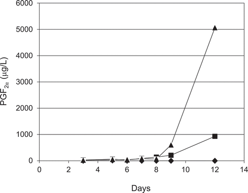
Figure 7. Time course analysis of extracellular PGF2α production in 12 days of cultivation by the parental (◆, M. alpina 1S-4) and transformant (■, GvMA#21 and ▲, GvMA#28) strains.