Figures & data
Scheme 1. The rapid competitive ELISA assay based on the epoxy-magnetic beads protocol.
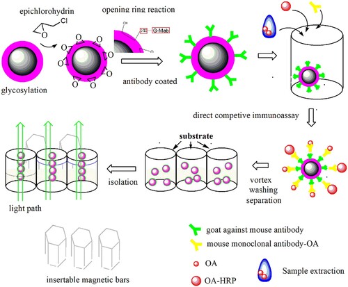
Figure 1. The morphology of bare magnetic beads and glycosylated beads, EDX profiles and Fourier transform infrared (FTIR) characterization (A) The SEM and EDX profiles of bare magnetic beads (B) The SEM and EDX profiles of glycosylated beads. (C) FTIR spectra of the glucose and glycosylated MBs samples: a-glucose b-glycosylated MBs.
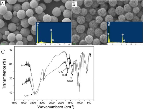
Figure 2. Cross-reactivity under the coexistence of other interferences. The IC50 investigation for OA ranged from 0.39 to 25.0 µg L−1, and DTX-1 ranged from 0.78 to 50.0 µg L−1. The assay condition of insert table consisted of the interfering substance at a concentration of 25.0 µg L−1.
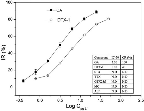
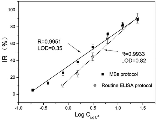
Figure 3. Calibration curves of the presented MB protocol, routine ELISA protocol.