2,260
Views
18
CrossRef citations to date
0
Altmetric
Articles

Silver nanoparticles from Prosopis glandulosa and their potential application as biocontrol of Acinetobacter calcoaceticus and Bacillus cereus

, , , , , & show all
Pages 1-5 | Received 25 Sep 2016, Accepted 21 Oct 2016, Published online: 02 Nov 2016

Figures & data

Figure 1. Synthesis of silver nanoparticles using plant extracts of P. glandulosa.

Figure 1. Synthesis of silver nanoparticles using plant extracts of P. glandulosa.

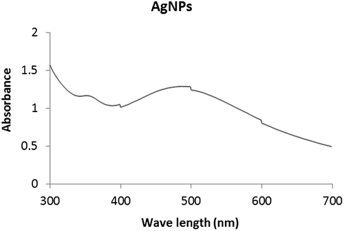
Figure 2. UV–Vis spectra of the synthesized silver nanoparticles.

Figure 2. UV–Vis spectra of the synthesized silver nanoparticles.

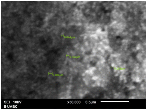
Figure 3. SEM image of silver nanoparticles from P. glandulosa.

Figure 3. SEM image of silver nanoparticles from P. glandulosa.

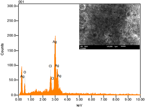
Figure 4. EDS image of silver nanoparticles produced from P. glandulosa.

Figure 4. EDS image of silver nanoparticles produced from P. glandulosa.

Table 1. Zeta-potential analysis of silver nanoparticles synthesized from P. glandulosa.

Figure 5. Particle size distribution of silver nanoparticles from dynamic light scattering measurements.
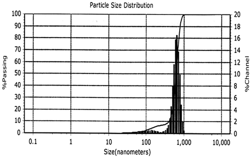
Figure 5. Particle size distribution of silver nanoparticles from dynamic light scattering measurements.

Figure 6. Antibacterial activity of P. glandulosa extract and AgNPs from P.glandulosa (a) Bacillus cereus and (b) Acinetobacter calcoaceticus.
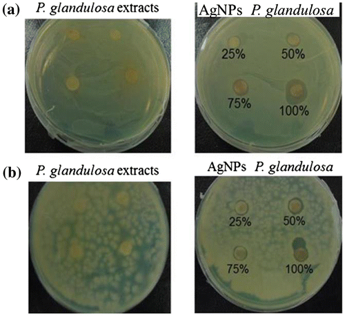
Figure 6. Antibacterial activity of P. glandulosa extract and AgNPs from P.glandulosa (a) Bacillus cereus and (b) Acinetobacter calcoaceticus.
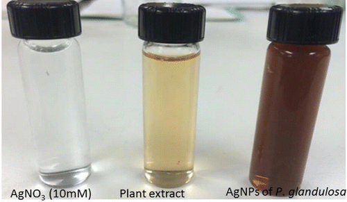
Table 2. Inhibition of microorganisms by AgNPs from P. glandulosa.