Figures & data
Fig. 1. (a) Light micrograph of Chlamydomonas nivalis cells with adhering material or mucilage sheet. (b) C. nivalis sample with smooth cell wall. (c) Snow algae cells, found in dry rock surface dust. These hypnoblasts were kept for 6 months in darkness and appeared viable after resuspension in water. (d) Sucrose density-gradient purification of C. nivalis concentrated from melting snow. The band formed in the middle of the gradient was used for most analyses. Scale bar: 10 µm
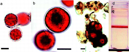
Table 1. Collection sites of ‘red snow’ algae in the Austrian Alps, province Tyrol. Chlamydomonas nivalis was always the dominant form. The snow from Sellrain/Kühtai, sampled in May, contained younger C. nivalis cell stages showing less accumulation of secondary carotenoids, and surface coating with organic and inorganic particles.
Fig. 2. Net photosynthesis vs. irradiance curves for snow algae at different temperatures. Values are means of three independent records, and error bars are standard deviations.
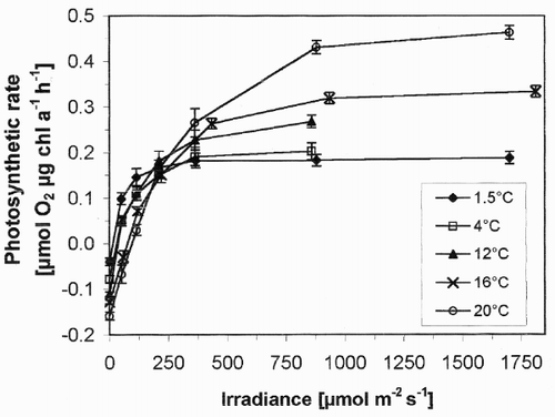
Table 2. Content of main pigments of snow algae sampled as in Table 1. Pigment contents are expressed in µg per µg chlorophyll a. Mean values calculated only from samples containing mostly adult cells; row 1 (spring harvest) not included. Replicate runs of each sample showed data variation of less than 1%. The description of sample plots is given in .
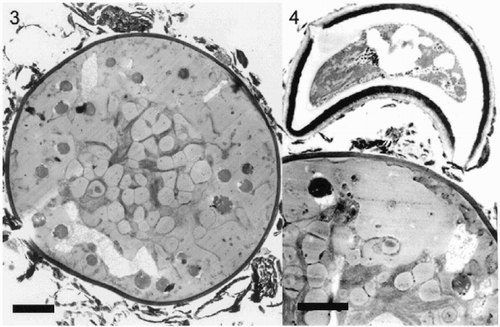
Figs 3–4. Ultrastructure of hypnoblasts of Chlamydomonas nivalis. Fig. 3. Typical older hypnoblast. The grey droplets are astaxanthin esters. In the central region, some plastid structures appear. Irregular structures of unknown composition, which were not removed by gradient separation, cover the outer cell wall. Scale bar: 2 µm. Fig. 4. Part of an older hypnoblast with adhering fungus-like structure. Scale bar: 2 µm.
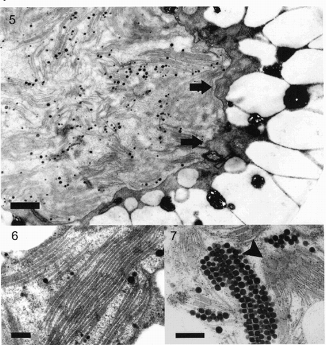
Figs 5–7. Fig. 5. Part of a Chlamydomonas nivalis cell showing the centrally located plastid surrounded by a thick layer of carotenoid globules with only a small cytoplasmic area left between both structures. The envelope membrane (arrows) of the plastid is undulating, the plastid contains numerous small grana stacks. Scale bar: 1 µm. Fig. 6. High magnification detail of thylakoid structure in Chlamydomonas nivalis cells showing a well-preserved ultrastructure. Such large stacks of thylakoids occur only occasionally. Scale bar: 0.2 µm. Fig. 7. Detail of plastid of Chlamydomonas nivalis with large numbers of plastoglobules, packed in regular arrangement. Some of the surrounding small grana stacks are visible in surface-view (arrowhead). Scale bar: 0.5 µm.