2,257
Views
19
CrossRef citations to date
0
Altmetric
Original Articles

Morphological and molecular characteristics of selected species of Minidiscus (Bacillariophyta, Thalassiosiraceae)

, , &
Pages 461-475 | Received 18 Jul 2008, Accepted 20 Feb 2009, Published online: 21 Aug 2009

Figures & data

Table 1.  Summary of Minidiscus culture codes, isolation dates and locations, GenBank and BOLD accession numbers.

Figs 1–6. Figs 1–2. Minidiscus chilensis. . Valve of a planktonic specimen with a thread (presumably chitin) extruded from a central fultoportula (short arrows) with a small external tube, a small external rimoportula opening (long arrow), and short, radial rows of simple pores (arrowhead). . Specimen from the benthos, sessile on a grain of sediment and covered with particles of clay; note the external fultoportula tubes (arrowheads). . Minidiscus comicus, all planktonic forms. . One individual with a thread (presumably chitin) extruding from one fultoportula with a large, wide open external tube (short arrows). . Recently divided cells with collapsed cingulum. Note the wide openings of the external fultoportula tubes (short arrows), one with a thread emerging. . A strongly silicified specimen with small areola openings (short arrows), wide open external fultoportula tubes and centrally located external rimoportula opening (long arrow). . Lightly (incompletely) silicified valve showing radial areola arrangement and the outlines of the bases of the fultoportula external tubes (arrowheads). Scale bars: 1 µm.
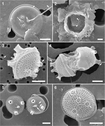
Figs 1–6. Figs 1–2. Minidiscus chilensis. Fig. 1. Valve of a planktonic specimen with a thread (presumably chitin) extruded from a central fultoportula (short arrows) with a small external tube, a small external rimoportula opening (long arrow), and short, radial rows of simple pores (arrowhead). Fig. 2. Specimen from the benthos, sessile on a grain of sediment and covered with particles of clay; note the external fultoportula tubes (arrowheads). Figs 3–6. Minidiscus comicus, all planktonic forms. Fig. 3. One individual with a thread (presumably chitin) extruding from one fultoportula with a large, wide open external tube (short arrows). Fig. 4. Recently divided cells with collapsed cingulum. Note the wide openings of the external fultoportula tubes (short arrows), one with a thread emerging. Fig. 5. A strongly silicified specimen with small areola openings (short arrows), wide open external fultoportula tubes and centrally located external rimoportula opening (long arrow). Fig. 6. Lightly (incompletely) silicified valve showing radial areola arrangement and the outlines of the bases of the fultoportula external tubes (arrowheads). Scale bars: 1 µm.

Table 2.  Morphometric summary of four strains of Minidiscusa .

Figs 7–10. Minidiscus trioculatus var. trioculatus, cultured material, CCMP strain 496 and a natural plankton specimen. . Larger, partially silicified, dome-like shaped valve, which possibly originated from an auxospore-like structure (see text for details); note characteristic wide basal part of the fultoportula external tube (short arrow). . Specimen from the natural environment, note tangential–linear valve face ornamentation and several copulae, one folded underneath the marginal flange of the valve (arrowhead). . An external view of a partially silicified valve with a tangential–linear areola pattern and a valvocopula with two rows of pores (arrowheads). Note the two satellite pores of the fultoportula (short arrows). . Internal valve view. Note the clusters of pores below the areolae (arrowheads), and the internal openings of two fultoportulae, each with two satellite pores (short arrows), and a rimoportula (long arrow). Scale bars: 1 µm.
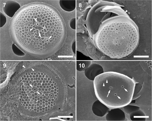
Figs 7–10. Minidiscus trioculatus var. trioculatus, cultured material, CCMP strain 496 and a natural plankton specimen. Fig. 7. Larger, partially silicified, dome-like shaped valve, which possibly originated from an auxospore-like structure (see text for details); note characteristic wide basal part of the fultoportula external tube (short arrow). Fig. 8. Specimen from the natural environment, note tangential–linear valve face ornamentation and several copulae, one folded underneath the marginal flange of the valve (arrowhead). Fig. 9. An external view of a partially silicified valve with a tangential–linear areola pattern and a valvocopula with two rows of pores (arrowheads). Note the two satellite pores of the fultoportula (short arrows). Fig. 10. Internal valve view. Note the clusters of pores below the areolae (arrowheads), and the internal openings of two fultoportulae, each with two satellite pores (short arrows), and a rimoportula (long arrow). Scale bars: 1 µm.

Figs 11–14. Minidiscus trioculatus var. monoculatus. . A typical specimen from a natural sample. Note one margin of the valvocopula is tucked underneath the marginal flange of the valve (short arrows). . Two valves from culture, showing a varying degree of areola development and the external openings of the fultoportula (arrowhead) and the rimoportula (long arrow). . A partially silicified valve illustrating tangential–linear areola organization and valvocopula with rows of pores (short arrow). . An internal view of a valve with a sub-central fultoportula (arrowhead), and a rimoportula (long arrow). Scale bars: 1 µm.
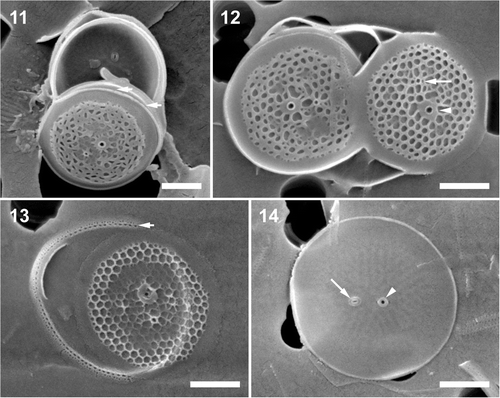
Figs 11–14. Minidiscus trioculatus var. monoculatus. Fig. 11. A typical specimen from a natural sample. Note one margin of the valvocopula is tucked underneath the marginal flange of the valve (short arrows). Fig. 12. Two valves from culture, showing a varying degree of areola development and the external openings of the fultoportula (arrowhead) and the rimoportula (long arrow). Fig. 13. A partially silicified valve illustrating tangential–linear areola organization and valvocopula with rows of pores (short arrow). Fig. 14. An internal view of a valve with a sub-central fultoportula (arrowhead), and a rimoportula (long arrow). Scale bars: 1 µm.

Figs 15–18. Minidiscus variabilis var. variabilis, cultured material, CCMP strain 495. . External valve view showing the typical, radial areola arrangement. . A frustule showing the areola distribution and the ornamentation of the valvocopula (short arrow). . An incompletely silicified valve showing the number and distribution of portulae. Note a number of radial ridges running from the valve near-centre to the marginal flange. . Internal view of radial rows of minute pores. Scale bars: 1 µm.
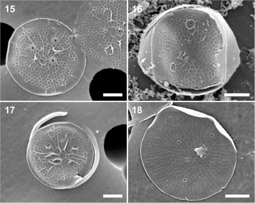
Figs 15–18. Minidiscus variabilis var. variabilis, cultured material, CCMP strain 495. Fig. 15. External valve view showing the typical, radial areola arrangement. Fig. 16. A frustule showing the areola distribution and the ornamentation of the valvocopula (short arrow). Fig. 17. An incompletely silicified valve showing the number and distribution of portulae. Note a number of radial ridges running from the valve near-centre to the marginal flange. Fig. 18. Internal view of radial rows of minute pores. Scale bars: 1 µm.

Figs 19–22. Scatter plots of the diameter and the number of fultoportulae in four, representative, sub-clones. There is no relationship between valve diameter and number of fultoportulae in CCMP 495. T1C7, T1B2, T1D3 and T1E1 indicate the individual subclones.
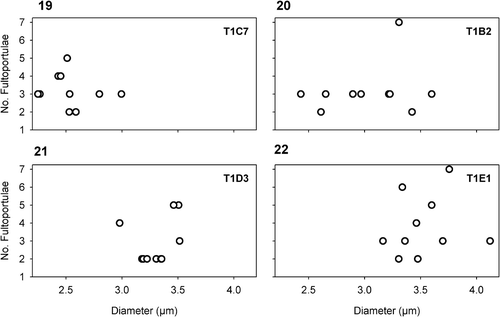
Figs 19–22. Scatter plots of the diameter and the number of fultoportulae in four, representative, sub-clones. There is no relationship between valve diameter and number of fultoportulae in CCMP 495. T1C7, T1B2, T1D3 and T1E1 indicate the individual subclones.

Figs 23–26. Minidiscus variabilis f. inornata. . A typically non-ornamented external view of a completely silicified valve showing a faint radial pattern with fultoportulae (arrowhead) and rimoportula (long arrow). Note that the fully formed external fultoportula tubes show that valve morphogenesis is complete. . Poroid areolae on copulae (short arrows). . External view of a valve with marginal ridges (short arrow) and a small external rimoportula opening (long arrow). . Internal view of a valve showing radiating rows of minute pores and the internal portula openings. Scale bars: 1 µm.
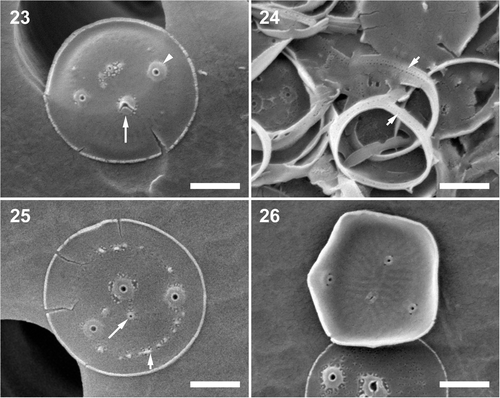
Figs 23–26. Minidiscus variabilis f. inornata. Fig. 23. A typically non-ornamented external view of a completely silicified valve showing a faint radial pattern with fultoportulae (arrowhead) and rimoportula (long arrow). Note that the fully formed external fultoportula tubes show that valve morphogenesis is complete. Fig. 24. Poroid areolae on copulae (short arrows). Fig. 25. External view of a valve with marginal ridges (short arrow) and a small external rimoportula opening (long arrow). Fig. 26. Internal view of a valve showing radiating rows of minute pores and the internal portula openings. Scale bars: 1 µm.

Fig. 27. A plot illustrating the relationship between valve diameter, number of areolae and fultoportula. Due to the frequent lack of perforations in M. variabilis f. inornata, it was only possible to quantify all characters in a small number of cells. Numbers in parentheses correspond to the number of fultoportulae per valve.
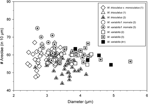
Fig. 27. A plot illustrating the relationship between valve diameter, number of areolae and fultoportula. Due to the frequent lack of perforations in M. variabilis f. inornata, it was only possible to quantify all characters in a small number of cells. Numbers in parentheses correspond to the number of fultoportulae per valve.

Table 3.  Comparison matrix of nucleotide differences within the ITS region of various Minidiscus species a .

Reprints and Corporate Permissions

Please note: Selecting permissions does not provide access to the full text of the article, please see our help page How do I view content?

To request a reprint or corporate permissions for this article, please click on the relevant link below:

Academic Permissions

Please note: Selecting permissions does not provide access to the full text of the article, please see our help page How do I view content?

Obtain permissions instantly via Rightslink by clicking on the button below:

If you are unable to obtain permissions via Rightslink, please complete and submit this Permissions form. For more information, please visit our Permissions help page.